

Pre-treatment with chemotherapy can enhance the antigenicity and immunogenicity of tumours by promoting adaptive immune responses
- PMID: 19997099
- PMCID: PMC2813751
- DOI: 10.1038/sj.bjc.6605465
Pre-treatment with chemotherapy can enhance the antigenicity and immunogenicity of tumours by promoting adaptive immune responses
Abstract
Background: Some cancer patients are immuno-compromised, and it has been long felt that immune-intervention is not compatible with standard chemotherapies. However, increasing evidence suggests that standard chemotherapy drugs may stimulate beneficial changes in both the immune system and tumour.
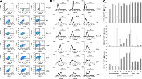
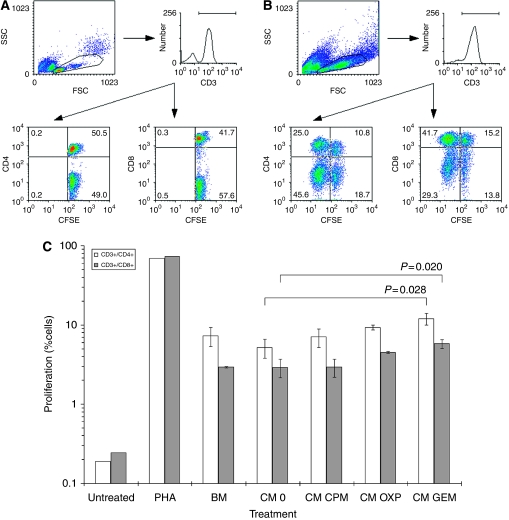
Methods: We have assessed the expression of human leucocyte antigen class 1 (HLA1) on tumour cells before and after chemotherapy agents (cyclophosphamide, oxaliplatin or gemcitabine). In addition, we show that chemotherapy-stressed tumour cells may release cytokines that enhance the interactions between dendritic cells (DCs) and T cells into growth media.
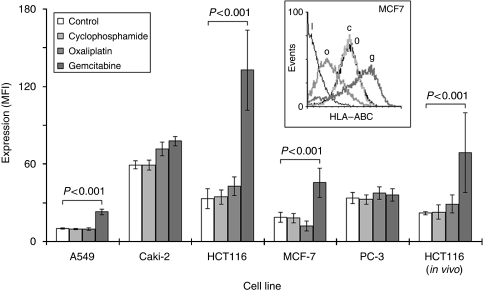
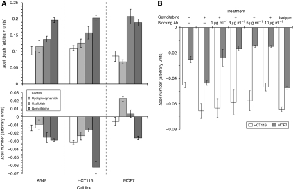
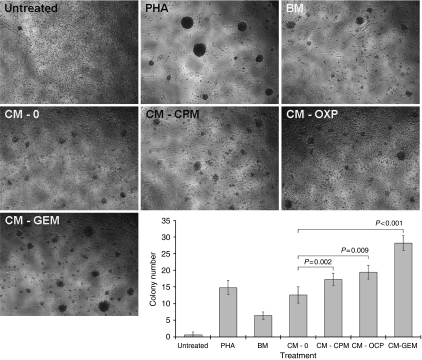
Results: Here we report that some chemotherapy agents can increase HLA1 expression in tumour cells, even when expression is low. Increases were associated with killing by cytotoxic T cells, which were negated by HLA1-blockade. Furthermore, T-cell function, as indicated by increased proliferation, was enhanced as supernatants derived from tumours treated with chemotherapy augmented DC-maturation and function.
Conclusion: There is evidence that a facet of immune surveillance can be restored by appropriate chemotherapy agents. Also, tumours exposed to some chemotherapy may secrete cytokines that can mature DCs, which ultimately enhances T-cell responses.
Figures
Similar articles
-
Immunological off-target effects of standard treatments in gastrointestinal cancers.Ann Oncol. 2014 Jan;25(1):24-32. doi: 10.1093/annonc/mdt349. Epub 2013 Nov 7. Ann Oncol. 2014. PMID: 24201974 Free PMC article. Review.
-
Dendritic cell-mediated cross-presentation of antigens derived from colon carcinoma cells exposed to a highly cytotoxic multidrug regimen with gemcitabine, oxaliplatin, 5-fluorouracil, and leucovorin, elicits a powerful human antigen-specific CTL response with antitumor activity in vitro.J Immunol. 2005 Jul 15;175(2):820-8. doi: 10.4049/jimmunol.175.2.820. J Immunol. 2005. PMID: 16002679
-
Supernatants from lymphocytes stimulated with Bacillus Calmette-Guerin can modify the antigenicity of tumours and stimulate allogeneic T-cell responses.Br J Cancer. 2011 Aug 23;105(5):687-93. doi: 10.1038/bjc.2011.306. Epub 2011 Aug 9. Br J Cancer. 2011. PMID: 21829193 Free PMC article.
-
Effect of Gemcitabine based chemotherapy on the immunogenicity of pancreatic tumour cells and T-cells.Clin Transl Oncol. 2021 Jan;23(1):110-121. doi: 10.1007/s12094-020-02429-0. Epub 2020 Jul 13. Clin Transl Oncol. 2021. PMID: 32661823 Free PMC article.
-
Adjuvants Enhancing Cross-Presentation by Dendritic Cells: The Key to More Effective Vaccines?Front Immunol. 2018 Dec 13;9:2874. doi: 10.3389/fimmu.2018.02874. eCollection 2018. Front Immunol. 2018. PMID: 30619259 Free PMC article. Review.
Cited by
-
Immunotherapy for pancreatic cancer: chasing the light at the end of the tunnel.Cell Oncol (Dordr). 2021 Apr;44(2):261-278. doi: 10.1007/s13402-021-00587-z. Epub 2021 Mar 12. Cell Oncol (Dordr). 2021. PMID: 33710604 Free PMC article. Review.
-
Cancer and HIV-1 Infection: Patterns of Chronic Antigen Exposure.Front Immunol. 2020 Jun 30;11:1350. doi: 10.3389/fimmu.2020.01350. eCollection 2020. Front Immunol. 2020. PMID: 32714330 Free PMC article. Review.
-
Pembrolizumab and salvage chemotherapy in EGFR T790M-positive non-small-cell lung cancer with high PD-L1 expression.Onco Targets Ther. 2018 Sep 7;11:5601-5605. doi: 10.2147/OTT.S168598. eCollection 2018. Onco Targets Ther. 2018. PMID: 30237726 Free PMC article.
-
Exploiting immune-dependent effects of microtubule-targeting agents to improve efficacy and tolerability of cancer treatment.Cell Death Dis. 2020 May 12;11(5):361. doi: 10.1038/s41419-020-2567-0. Cell Death Dis. 2020. PMID: 32398657 Free PMC article. Review.
-
Immunological off-target effects of standard treatments in gastrointestinal cancers.Ann Oncol. 2014 Jan;25(1):24-32. doi: 10.1093/annonc/mdt349. Epub 2013 Nov 7. Ann Oncol. 2014. PMID: 24201974 Free PMC article. Review.
References
-
- Aptsiauri N, Cabrera T, Mendez R, Garcia-Lora A, Ruiz-Cabello F, Garrido F (2007) Role of altered expression of HLA class I molecules in cancer progression. Adv Exp Med Biol 601: 123–131 - PubMed
-
- Berd D, Maguire Jr HC, Mastrangelo MJ (1984) Potentiation of human cell-mediated and humoral immunity by low-dose cyclophosphamide. Cancer Res 44: 5439–5443 - PubMed
-
- Bianco R, Melisi D, Ciardiello F, Tortora G (2006) Key cancer cell signal transduction pathways as therapeutic targets. Eur J Cancer 42: 290–294 - PubMed
-
- Brode S, Cooke A (2008) Immune-potentiating effects of the chemotherapeutic drug cyclophosphamide. Crit Rev Immunol 28: 109–126 - PubMed
Publication types
MeSH terms
Substances
LinkOut - more resources
Full Text Sources
Other Literature Sources
