

Secreted frizzled-related protein disrupts PCP in eye lens fiber cells that have polarised primary cilia
- PMID: 19968984
- PMCID: PMC2815166
- DOI: 10.1016/j.ydbio.2009.11.033
Secreted frizzled-related protein disrupts PCP in eye lens fiber cells that have polarised primary cilia
Abstract
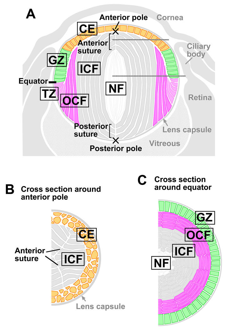
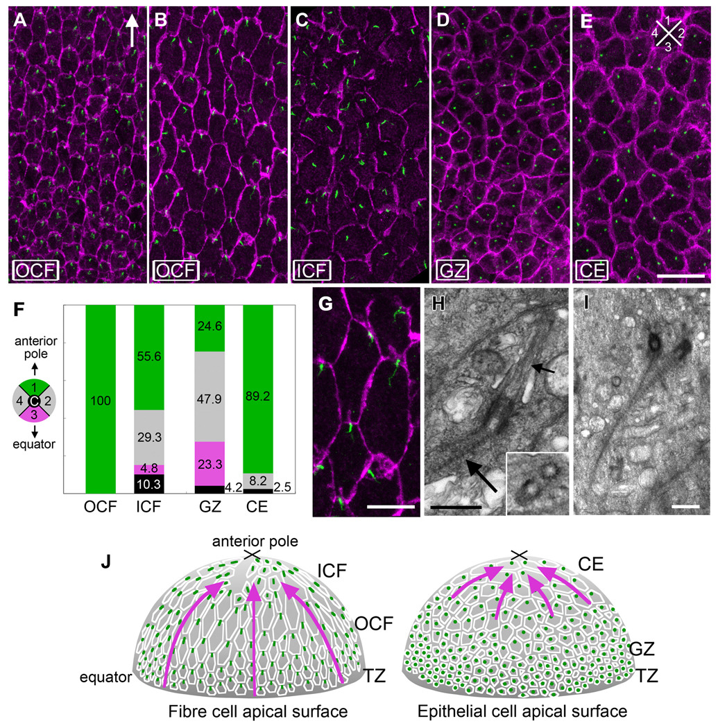
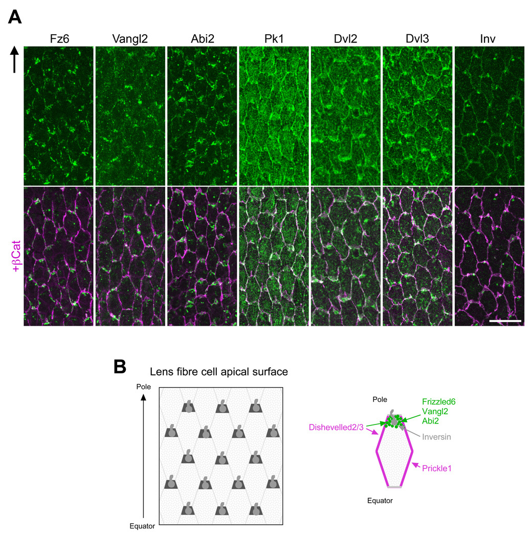
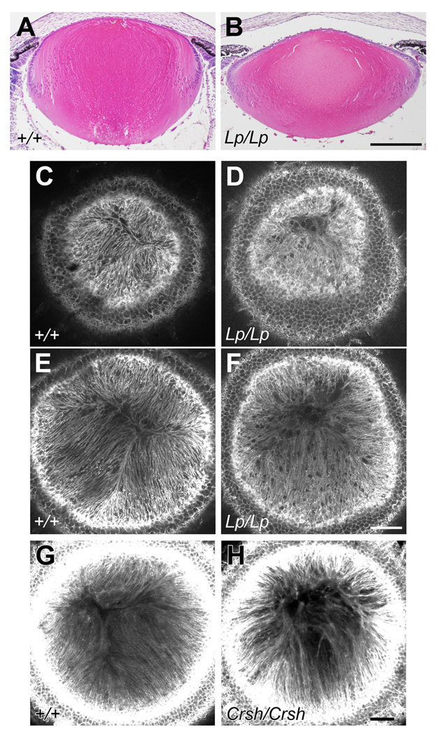
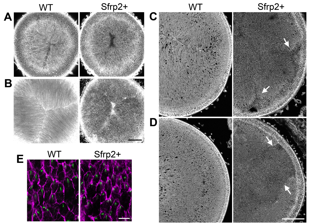
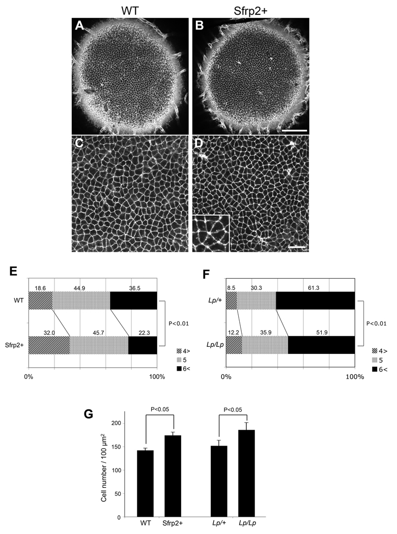
Planar cell polarity (PCP) signaling polarises cells along tissue axes. Although pathways involved are becoming better understood, outstanding issues include; (i) existence/identity of cues that orchestrate global polarisation in tissues, and (ii) the generality of the link between polarisation of primary cilia and asymmetric localisation of PCP proteins. Mammalian lenses are mainly comprised of epithelial-derived fiber cells. Concentrically arranged fibers are precisely aligned as they elongate along the anterior-posterior axis and orientate towards lens poles where they meet fibers from other segments to form characteristic sutures. We show that lens exhibits PCP, with each fiber cell having an apically situated cilium and in most cases this is polarised towards the anterior pole. Frizzled and other PCP proteins are also asymmetrically localised along the equatorial-anterior axis. Mutations in core PCP genes Van Gogh-like 2 and Celsr1 perturb oriented fiber alignment and suture formation. Suppression of the PCP pathway by overexpressing Sfrp2 shows that whilst local groups of fibers are often similarly oriented, they lack global orientation; consequently when local groups of fibers with different orientations meet they form multiple, small, ectopic suture-like configurations. This indicates that this extracellular inhibitor disrupts a global polarising signal that utilises a PCP-mediated mechanism to coordinate the global alignment and orientation of fibers to lens poles.
Copyright 2009 Elsevier Inc. All rights reserved.
Figures
Similar articles
-
Non-essential role for cilia in coordinating precise alignment of lens fibres.Mech Dev. 2016 Feb;139:10-7. doi: 10.1016/j.mod.2016.01.003. Epub 2016 Jan 26. Mech Dev. 2016. PMID: 26825015 Free PMC article.
-
Analysis of PCP defects in mammalian eye lens.Methods Mol Biol. 2012;839:147-56. doi: 10.1007/978-1-61779-510-7_12. Methods Mol Biol. 2012. PMID: 22218899 Free PMC article.
-
A conserved function for Strabismus in establishing planar cell polarity in the ciliated ectoderm during cnidarian larval development.Development. 2012 Dec 1;139(23):4374-82. doi: 10.1242/dev.084251. Epub 2012 Oct 24. Development. 2012. PMID: 23095884
-
Planar cell polarity in the mammalian eye lens.Organogenesis. 2011 Jul-Sep;7(3):191-201. doi: 10.4161/org.7.3.18421. Epub 2011 Jul 1. Organogenesis. 2011. PMID: 22027540 Free PMC article. Review.
-
Frizzled/PCP signalling: a conserved mechanism regulating cell polarity and directed motility.Nat Rev Genet. 2007 Feb;8(2):126-38. doi: 10.1038/nrg2042. Nat Rev Genet. 2007. PMID: 17230199 Review.
Cited by
-
Analysis of genome-wide knockout mouse database identifies candidate ciliopathy genes.Sci Rep. 2022 Dec 1;12(1):20791. doi: 10.1038/s41598-022-19710-7. Sci Rep. 2022. PMID: 36456625 Free PMC article.
-
Planar cell polarity signaling in the Drosophila eye.Curr Top Dev Biol. 2010;93:189-227. doi: 10.1016/B978-0-12-385044-7.00007-2. Curr Top Dev Biol. 2010. PMID: 20959167 Free PMC article. Review.
-
Interactions between lens epithelial and fiber cells reveal an intrinsic self-assembly mechanism.Dev Biol. 2014 Jan 15;385(2):291-303. doi: 10.1016/j.ydbio.2013.10.030. Epub 2013 Nov 8. Dev Biol. 2014. PMID: 24211762 Free PMC article.
-
Fibrosis in the lens. Sprouty regulation of TGFβ-signaling prevents lens EMT leading to cataract.Exp Eye Res. 2016 Jan;142:92-101. doi: 10.1016/j.exer.2015.02.004. Epub 2015 May 21. Exp Eye Res. 2016. PMID: 26003864 Free PMC article. Review.
-
Diverse roles of Eph/ephrin signaling in the mouse lens.PLoS One. 2011;6(11):e28147. doi: 10.1371/journal.pone.0028147. Epub 2011 Nov 29. PLoS One. 2011. PMID: 22140528 Free PMC article.
References
-
- Axelrod JD. Basal bodies, kinocilia and planar cell polarity. Nat. Genet. 2008;40:10–11. - PubMed
-
- Benzing T, Walz G. Cilium-generated signaling: a cellular GPS? Curr. Opin. Nephrol. Hypertens. 2006;15:245–249. - PubMed
-
- Bovolenta P, Esteve P, Ruiz JM, Cisneros E, Lopez-Rios J. Beyond Wnt inhibition: new functions of secreted Frizzled-related proteins in development and disease. J. Cell Sci. 2008;121:737–746. - PubMed
-
- Classen AK, Anderson KI, Marois E, Eaton S. Hexagonal packing of Drosophila wing epithelial cells by the planar cell polarity pathway. Dev. Cell. 2005;9:805–817. - PubMed
Publication types
MeSH terms
Substances
Grants and funding
LinkOut - more resources
Full Text Sources
Other Literature Sources
Miscellaneous
