Nuclear receptor-induced chromosomal proximity and DNA breaks underlie specific translocations in cancer
- PMID: 19962179
- PMCID: PMC2812435
- DOI: 10.1016/j.cell.2009.11.030
Nuclear receptor-induced chromosomal proximity and DNA breaks underlie specific translocations in cancer
Abstract
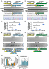
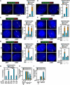
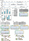
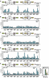
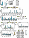
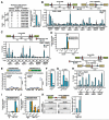
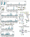
Chromosomal translocations are a hallmark of leukemia/lymphoma and also appear in solid tumors, but the underlying mechanism remains elusive. By establishing a cellular model that mimics the relative frequency of authentic translocation events without proliferation selection, we report mechanisms of nuclear receptor-dependent tumor translocations. Intronic binding of liganded androgen receptor (AR) first juxtaposes translocation loci by triggering intra- and interchromosomal interactions. AR then promotes site-specific DNA double-stranded breaks (DSBs) at translocation loci by recruiting two types of enzymatic activities induced by genotoxic stress and liganded AR, including activation-induced cytidine deaminase and the LINE-1 repeat-encoded ORF2 endonuclease. These enzymes synergistically generate site-selective DSBs at juxtaposed translocation loci that are ligated by nonhomologous end joining pathway for specific translocations. Our data suggest that the confluence of two parallel pathways initiated by liganded nuclear receptor and genotoxic stress underlies nonrandom tumor translocations, which may function in many types of tumors and pathological processes.
Figures
Comment in
-
The dangers of transcription.Cell. 2009 Dec 11;139(6):1047-9. doi: 10.1016/j.cell.2009.11.037. Cell. 2009. PMID: 20005797 Free PMC article.
Similar articles
-
NKX3.1 Suppresses TMPRSS2-ERG Gene Rearrangement and Mediates Repair of Androgen Receptor-Induced DNA Damage.Cancer Res. 2015 Jul 1;75(13):2686-98. doi: 10.1158/0008-5472.CAN-14-3387. Epub 2015 May 14. Cancer Res. 2015. PMID: 25977336 Free PMC article.
-
TMPRSS2:ERG blocks neuroendocrine and luminal cell differentiation to maintain prostate cancer proliferation.Oncogene. 2015 Jul;34(29):3815-25. doi: 10.1038/onc.2014.308. Epub 2014 Sep 29. Oncogene. 2015. PMID: 25263440
-
TMPRSS2:ERG fusion by translocation or interstitial deletion is highly relevant in androgen-dependent prostate cancer, but is bypassed in late-stage androgen receptor-negative prostate cancer.Cancer Res. 2006 Nov 15;66(22):10658-63. doi: 10.1158/0008-5472.CAN-06-1871. Cancer Res. 2006. PMID: 17108102
-
Mechanisms that promote and suppress chromosomal translocations in lymphocytes.Annu Rev Immunol. 2011;29:319-50. doi: 10.1146/annurev-immunol-031210-101329. Annu Rev Immunol. 2011. PMID: 21219174 Review.
-
Mechanisms of oncogenic chromosomal translocations.Ann N Y Acad Sci. 2014 Mar;1310:89-97. doi: 10.1111/nyas.12370. Epub 2014 Feb 16. Ann N Y Acad Sci. 2014. PMID: 24528169 Review.
Cited by
-
5α-reductase inhibitors impact prognosis of urothelial carcinoma.BMC Cancer. 2020 Sep 11;20(1):872. doi: 10.1186/s12885-020-07373-4. BMC Cancer. 2020. PMID: 32917158 Free PMC article.
-
Frequency of close positioning of chromosomal loci detected by FRET correlates with their participation in carcinogenic rearrangements in human cells.Genes Chromosomes Cancer. 2012 Nov;51(11):1037-44. doi: 10.1002/gcc.21988. Epub 2012 Aug 10. Genes Chromosomes Cancer. 2012. PMID: 22887574 Free PMC article.
-
Factors That Affect the Formation of Chromosomal Translocations in Cells.Cancers (Basel). 2022 Oct 18;14(20):5110. doi: 10.3390/cancers14205110. Cancers (Basel). 2022. PMID: 36291894 Free PMC article. Review.
-
The role of mechanistic factors in promoting chromosomal translocations found in lymphoid and other cancers.Adv Immunol. 2010;106:93-133. doi: 10.1016/S0065-2776(10)06004-9. Adv Immunol. 2010. PMID: 20728025 Free PMC article. Review.
-
Anakoinosis: Correcting Aberrant Homeostasis of Cancer Tissue-Going Beyond Apoptosis Induction.Front Oncol. 2019 Dec 20;9:1408. doi: 10.3389/fonc.2019.01408. eCollection 2019. Front Oncol. 2019. PMID: 31921665 Free PMC article. Review.
References
-
- Aguilera A, Gomez-Gonzalez B. Genome instability: a mechanistic view of its causes and consequences. Nat Rev Genet. 2008;9:204–217. - PubMed
-
- Aravin AA, Sachidanandam R, Girard A, Fejes-Toth K, Hannon GJ. Science. Vol. 316. New York, NY: 2007. Developmentally regulated piRNA clusters implicate MILI in transposon control; pp. 744–747. - PubMed
-
- Berkovich E, Monnat RJ, Jr., Kastan MB. Assessment of protein dynamics and DNA repair following generation of DNA double-strand breaks at defined genomic sites. Nature protocols. 2008;3:915–922. - PubMed
Publication types
MeSH terms
Substances
Grants and funding
- R01 NS034934/NS/NINDS NIH HHS/United States
- R37 DK039949-26/DK/NIDDK NIH HHS/United States
- R37 DK039949/DK/NIDDK NIH HHS/United States
- KG080247/PHS HHS/United States
- DK18477/DK/NIDDK NIH HHS/United States
- R01 CA097134-07/CA/NCI NIH HHS/United States
- R01 CA097134/CA/NCI NIH HHS/United States
- R01 NS034934-19/NS/NINDS NIH HHS/United States
- CA97134/CA/NCI NIH HHS/United States
- R01 HL065445-10/HL/NHLBI NIH HHS/United States
- R01 HL065445/HL/NHLBI NIH HHS/United States
- DK39949/DK/NIDDK NIH HHS/United States
- NS34934/NS/NINDS NIH HHS/United States
- HHMI/Howard Hughes Medical Institute/United States
LinkOut - more resources
Full Text Sources
Other Literature Sources
Medical
Research Materials

