

Internal lipid synthesis and vesicle growth as a step toward self-reproduction of the minimal cell
- PMID: 19957048
- PMCID: PMC2923298
- DOI: 10.1007/s11693-009-9048-1
Internal lipid synthesis and vesicle growth as a step toward self-reproduction of the minimal cell
Abstract
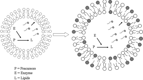
One of the major properties of the semi-synthetic minimal cell, as a model for early living cells, is the ability to self-reproduce itself, and the reproduction of the boundary layer or vesicle compartment is part of this process. A minimal bio-molecular mechanism based on the activity of one single enzyme, the FAS-B (Fatty Acid Synthase) Type I enzyme from Brevibacterium ammoniagenes, is encapsulated in 1-palmitoyl-2oleoyl-sn-glycero-3-phosphatidylcholine (POPC) liposomes to control lipid synthesis. Consequently molecules of palmitic acid released from the FAS catalysis, within the internal lumen, move toward the membrane compartment and become incorporated into the phospholipid bilayer. As a result the vesicle membranes change in lipid composition and liposome growth can be monitored. Here we report the first experiments showing vesicles growth by catalysis of one enzyme only that produces cell boundary from within. This is the prototype of the simplest autopoietic minimal cell.
Figures
Similar articles
-
Early self-reproduction, the emergence of division mechanisms in protocells.Mol Biosyst. 2013 Feb 2;9(2):195-204. doi: 10.1039/c2mb25375e. Epub 2012 Dec 11. Mol Biosyst. 2013. PMID: 23232904 Review.
-
Kinetic studies of the interaction of fatty acids with phosphatidylcholine vesicles (liposomes).Colloids Surf B Biointerfaces. 2006 Mar 1;48(1):24-34. doi: 10.1016/j.colsurfb.2006.01.001. Epub 2006 Feb 8. Colloids Surf B Biointerfaces. 2006. PMID: 16466910
-
Question 7: construction of a semi-synthetic minimal cell: a model for early living cells.Orig Life Evol Biosph. 2007 Oct;37(4-5):419-22. doi: 10.1007/s11084-007-9090-5. Epub 2007 Aug 1. Orig Life Evol Biosph. 2007. PMID: 17668286
-
Effects of specific fatty acid acylation of phospholipase A2 on its interfacial binding and catalysis.Biochemistry. 1994 Sep 27;33(38):11598-607. doi: 10.1021/bi00204a022. Biochemistry. 1994. PMID: 7918373
-
Enzymes inside lipid vesicles: preparation, reactivity and applications.Biomol Eng. 2001 Oct 31;18(4):143-77. doi: 10.1016/s1389-0344(01)00088-0. Biomol Eng. 2001. PMID: 11576871 Review.
Cited by
-
Protocells programmed through artificial reaction networks.Chem Sci. 2019 Dec 19;11(3):631-642. doi: 10.1039/c9sc05043d. Chem Sci. 2019. PMID: 34123035 Free PMC article. Review.
-
Synthetic Minimal Cell: Self-Reproduction of the Boundary Layer.ACS Omega. 2019 Mar 31;4(3):5293-5303. doi: 10.1021/acsomega.8b02955. Epub 2019 Mar 13. ACS Omega. 2019. PMID: 30949617 Free PMC article.
-
Genetically controlled membrane synthesis in liposomes.Nat Commun. 2020 Aug 28;11(1):4317. doi: 10.1038/s41467-020-17863-5. Nat Commun. 2020. PMID: 32859896 Free PMC article.
-
Divided we stand: splitting synthetic cells for their proliferation.Syst Synth Biol. 2014 Sep;8(3):249-69. doi: 10.1007/s11693-014-9145-7. Epub 2014 May 27. Syst Synth Biol. 2014. PMID: 25136387 Free PMC article. Review.
-
Vesicle-based artificial cells: materials, construction methods and applications.Mater Horiz. 2022 Mar 7;9(3):892-907. doi: 10.1039/d1mh01431e. Mater Horiz. 2022. PMID: 34908080 Free PMC article. Review.
References
-
- Bachmann PA, Luisi PL, Lang J. Autocatalytic self-replicating micelles as models for prebiotic structures. Nature. 1992;357:57–59. doi: 10.1038/357057a0. - DOI
-
- Berclaz N, Muller M, Walde P, Luisi PL. Growth and transformation of vesicles studied by ferritin labelling and cryotransmission electron microscopy. J Phys Chem B. 2001;105:1056–1064. doi: 10.1021/jp001298i. - DOI
LinkOut - more resources
Full Text Sources
Research Materials
Miscellaneous
