. 2010 Feb;84(4):2110-21.
doi: 10.1128/JVI.02007-09. Epub 2009 Dec 2.

Nucleolin is required for efficient nuclear egress of herpes simplex virus type 1 nucleocapsids

Affiliations

Nucleolin is required for efficient nuclear egress of herpes simplex virus type 1 nucleocapsids

Ken Sagou et al. J Virol. 2010 Feb.

Abstract

Herpesvirus nucleocapsids assemble in the nucleus and must cross the nuclear membrane for final assembly and maturation to form infectious progeny virions in the cytoplasm. It has been proposed that nucleocapsids enter the perinuclear space by budding through the inner nuclear membrane, and these enveloped nucleocapsids then fuse with the outer nuclear membrane to enter the cytoplasm. Little is known about the mechanism(s) for nuclear egress of herpesvirus nucleocapsids and, in particular, which, if any, cellular proteins are involved in the nuclear egress pathway. UL12 is an alkaline nuclease encoded by herpes simplex virus type 1 (HSV-1) and has been suggested to be involved in viral DNA maturation and nuclear egress of nucleocapsids. Using a live-cell imaging system to study cells infected by a recombinant HSV-1 expressing UL12 fused to a fluorescent protein, we observed the previously unreported nucleolar localization of UL12 in live infected cells and, using coimmunoprecipitation analyses, showed that UL12 formed a complex with nucleolin, a nucleolus marker, in infected cells. Knockdown of nucleolin in HSV-1-infected cells reduced capsid accumulation, as well as the amount of viral DNA resistant to staphylococcal nuclease in the cytoplasm, which represented encapsidated viral DNA, but had little effect on these viral components in the nucleus. These results indicated that nucleolin is a cellular factor required for efficient nuclear egress of HSV-1 nucleocapsids in infected cells.


Figures
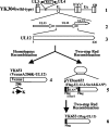
FIG. 1.
FIG. 1.
Schematic diagram of genome structure of wild-type YK304 and relevant recombinant virus domains. Line 1, linear representation of the YK304 genome. The YK304 genome has a bacmid (BAC) in the intergenic region between UL3 and UL4. Line 2, fragment of the genome domain containing the UL11, UL12, and UL13 ORFs. Line 3, UL12 ORF. Lines 4 and 6, recombinant viruses YK651 and YK653, respectively. Line 5, plasmid pYK652.
FIG. 2.
FIG. 2.
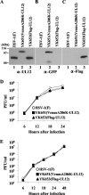
Characterization of recombinant viruses. (A to C) Immunoblots of electrophoretically separated lysates from Vero cells infected with wild-type HSV-1(F) (lane 1), YK651 (Venus-UL12) (lane 2), and YK653 (Flag-UL12) (lane 3). Infected cells were harvested 18 h postinfection and analyzed by immunoblotting with antibody to UL12 (A), GFP (B), and Flag (C). (D and E) Vero cells were infected at an MOI of 5 (D) or 0.01 (E) with wild-type HSV-1(F), YK651 (Venus-UL12), or YK653 (Flag-UL12). Total virus from cell culture supernatants and infected cells harvested at the indicated times were assayed on Vero cells.
FIG. 3.
FIG. 3.
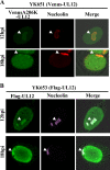
Localization of nucleolin and UL12 in infected cells. Vero cells infected with YK651 (Venus-UL12) (A) or YK653 (Flag-UL12) (B) were fixed at 12 and 18 h postinfection and permeabilized. YK651-infected cells were then treated with antibody to nucleolin, which was detected with Alexa Fluor 546-conjugated donkey anti-mouse IgG by confocal microscopy. YK653-infected cells were then treated with antibodies to Flag and nucleolin, which were detected with Alexa Fluor 488-conjugated goat anti-rabbit IgG and Alexa Fluor 680-conjugated goat anti-mouse IgG, respectively, by confocal microscopy. The triangles indicate nuclear compartments that appeared to be nucleoli.
FIG. 4.
FIG. 4.
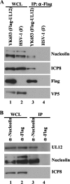
Identification of the UL12-nucleolin complex. (A) Vero cells infected with YK653 (Flag-UL12) (lanes 1 and 3) or wild-type HSV-1(F) (lanes 2 and 4) were lysed and immunoprecipitated with antibody to the Flag epitope. The immunoprecipitates (lanes 3 and 4) were analyzed by electrophoresis and immunoblotted with antibody to nucleolin, ICP8, Flag, and VP5. One-ninth of the Vero whole-cell lysates (WCL) used in the reaction mixtures for lanes 3 and 4 was loaded in lanes 1 and 2, respectively. (B) Vero cells infected with wild-type HSV-1(F) were lysed and immunoprecipitated with antibody to nucleolin (lanes 1 and 3) and Flag (lanes 2 and 4). The immunoprecipitates (lanes 3 and 4) were analyzed by electrophoresis and immunoblotted with antibody to UL12, ICP8, and nucleolin. One-ninth of the Vero WCLs used in the reaction mixtures for lanes 3 and 4 was loaded in lanes 1 and 2, respectively.
FIG. 5.
FIG. 5.
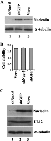
Knockdown of nucleolin. (A) Vero-shNuc-5 cells expressing shRNA against nucleolin (lane 1), Vero-shGFP cells expressing shRNA against GFP (lane 2), and normal Vero cells (lane 3) were analyzed by immunoblotting with antibody to nucleolin (upper panel) and α-tubulin (lower panel). (B) The viability of Vero, Vero-shNuc-5, and Vero-shGFP cells relative to a control (medium only) was measured as the cell metabolic activity. Each value is the mean ± the standard deviation of three independent experiments. (C) Vero-shNuc-5, Vero-shGFP, and Vero cells infected with wild-type HSV-1(F) for 12 h were analyzed by immunoblotting with antibody to nucleolin, UL12, and α-tubulin.
FIG. 6.
FIG. 6.
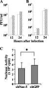
Effect of nucleolin knockdown on viral growth and nuclease activity. (A and B) Vero-shNuc-5 (open bar) and Vero-shGFP (shaded bar) cells were infected with HSV-1(F) at an MOI of 5 (A) or 0.01 (B). At 12 and 24 h postinfection, total virus from cell culture supernatants and infected cells was harvested and assayed on Vero cells. (C) Alkaline nuclease activity in HSV-1(F)-infected Vero-shNuc-5 and Vero-shGFP cells. Nuclease activity in each cell line was calculated by subtracting the activity in mock-infected cells from that in cells infected with HSV-1(F). Each value is the mean ± the standard deviation of three independent experiments. The statistical difference of the nuclease activity between Vero-shNuc- and Vero-shGFP-infected cells is noted (*, P < 0.05).
FIG. 7.
FIG. 7.
SN sensitivity analysis of HSV-1 DNA during viral replication. (A) Vero-shNuc-5 (lanes 1 and 3) and Vero-shGFP (lanes 2 and 4) cells infected with wild-type HSV-1(F) at an MOI of 5 were fractionated into cytoplasmic (lanes 1 and 2) and nuclear (lanes 3 and 4) fractions at 12 h postinfection, and the fractions were treated with SN. DNA from each fraction and purified virion DNA (lane 5) were analyzed by Southern blotting with UL41 DNA as the probe. (B) The amount of SN-resistant viral DNA in the cytoplasmic fractions was quantitated relative to the amount in the nuclear fractions from the data in panel A. The shGFP value was normalized to 100. (C) Vero-shNuc-5 (lanes 1 and 3) and Vero-shGFP (lanes 2 and 4) cells infected with wild-type HSV-1(F) were fractionated into cytoplasmic (lanes 1 and 2) and nuclear fractions (lanes 3 and 4) as described in panel A and were analyzed by Southern blotting with UL41 DNA as the probe without SN digestion. (D) The amount of viral DNA in the cytoplasmic fractions was quantitated relative to the amount of total viral DNA in the nuclear fractions from the data in panel C. The shGFP value was normalized to 100.
FIG. 8.
FIG. 8.
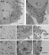
Election microscopy of infected nucleolin knockdown cells. Electron microscopy of wild-type HSV-1(F)-infected Vero-shGFP (A) and Vero-shNuc-5 cells (B). Panels a to f show magnifications of the corresponding areas indicated in panels A and B. Scale bar: A, 700 nm; B, 1,000 nm; a to f, 500 nm.
FIG. 9.
FIG. 9.
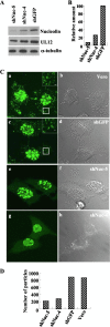
Confocal microscopy of infected nucleolin knock-down cells. (A) Vero-shNuc-4, Vero-shNuc-5, and Vero-shGFP cells infected with wild-type HSV-1(F) for 12 h were analyzed by immunoblotting with antibodies to nucleolin, UL12, and α-tubulin. (B) Quantitation of the amount of nucleolin protein relative to the amount of α-tubulin shown in panel A. The shGFP value was normalized to 100. (C) Vero (a and b), Vero-shGFP (c and d), Vero-shNuc-5 (e and f), and Vero-shNuc-4 (g and h) cells were infected with YK601 (VenusA206K-VP26) for 12 h and then examined by confocal micros- copy. Confocal Z-sections were acquired through the entire thickness of the cells. A confocal Z-stacked fluorescence image derived from the Z-sections (a, c, e, and g) and DIC (b, d, f, and h) images is shown. Insets show magnified images of the boxed areas. Scale bar, 500 nm. (D) Quantitation of virus particles in the cytoplasm of infected cells from the data in panel C.

Similar articles

Cited by

References

    1. Bjerke, S. L., and R. J. Roller. 2006. Roles for herpes simplex virus type 1 UL34 and US3 proteins in disrupting the nuclear lamina during herpes simplex virus type 1 egress. Virology 347:261-276. - PMC - PubMed
    1. Calle, A., I. Ugrinova, A. L. Epstein, P. Bouvet, J. J. Diaz, and A. Greco. 2008. Nucleolin is required for an efficient herpes simplex virus type 1 infection. J. Virol. 82:4762-4773. - PMC - PubMed
    1. Campadelli-Fiume, G., and B. Roizman. 2006. The egress of herpesviruses from cells: the unanswered questions. J. Virol. 80:6716-6719. - PMC - PubMed
    1. De, A., S. L. Donahue, A. Tabah, N. E. Castro, N. Mraz, J. L. Cruise, and C. Campbell. 2006. A novel interaction of nucleolin with Rad51. Biochem. Biophys. Res. Commun. 344:206-213. - PubMed
    1. Elliott, G., and P. O'Hare. 1999. Live-cell analysis of a green fluorescent protein-tagged herpes simplex virus infection. J. Virol. 73:4110-4119. - PMC - PubMed

Publication types

MeSH terms

LinkOut - more resources