. 2009 Nov 25;16(11):1158-68.
doi: 10.1016/j.chembiol.2009.10.006.

Identification and characterization of a small molecule inhibitor of formin-mediated actin assembly

Affiliations

Identification and characterization of a small molecule inhibitor of formin-mediated actin assembly

Syed A Rizvi et al. Chem Biol. .

Abstract

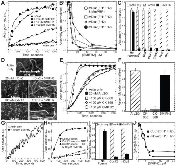
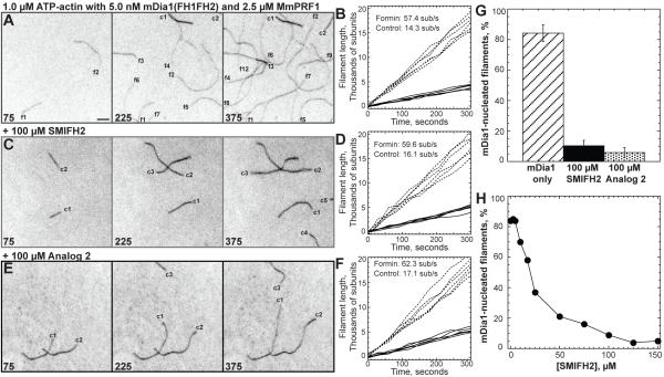
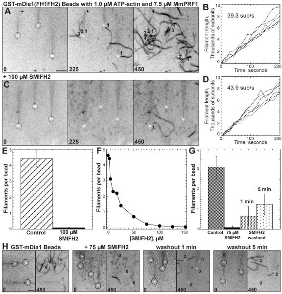
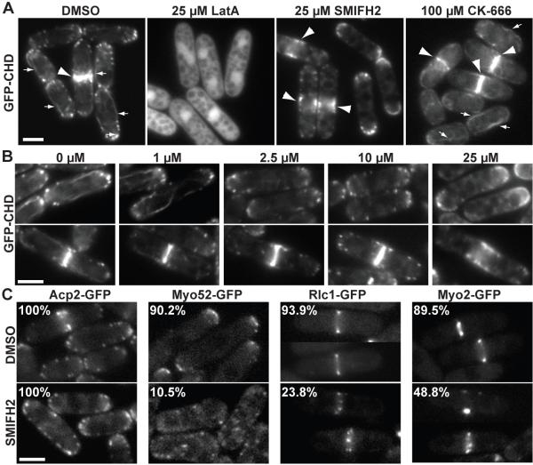
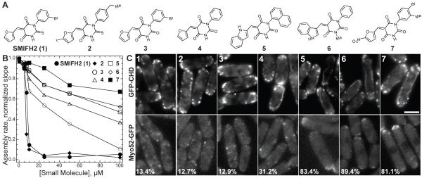
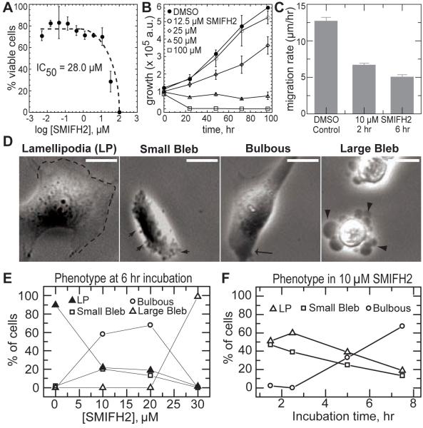
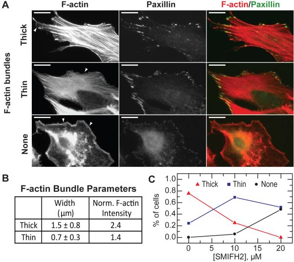
Formins stimulate actin filament assembly for fundamental cellular processes including division, adhesion, establishing polarity, and motility. A formin inhibitor would be useful because most cells express multiple formins whose functions are not known and because metastatic tumor formation depends on the deregulation of formin-dependent processes. We identified a general small molecule inhibitor of formin homology 2 domains (SMIFH2) by screening compounds for the ability to prevent formin-mediated actin assembly in vitro. SMIFH2 targets formins from evolutionarily diverse organisms including yeast, nematode worm, and mice, with a half-maximal inhibitor concentration of approximately 5 to 15 microM. SMIFH2 prevents both formin nucleation and processive barbed end elongation and decreases formin's affinity for the barbed end. Furthermore, low micromolar concentrations of SMIFH2 disrupt formin-dependent, but not Arp2/3 complex-dependent, actin cytoskeletal structures in fission yeast and mammalian NIH 3T3 fibroblasts.


Figures

Figure 1
Figure 1. SMIFH2 Inhibits Formin-Mediated Actin Assembly In Vitro
(A) Time course of the polymerization of 2.5 μM Mg-ATP actin monomers (20% pyrene-labeled) with 2.5 μM profilin MmPRF1 in either the absence (thick curve) or presence of 25 nM mDia1(FH1FH2) and 0.0 (■ ), 7.5 (● ), 10 (◆ ), or 100 μM SMIFH2 (▲ ). Conditions: 10 mM imidazole, pH 7.0, 50 mM KCl, 1 mM MgCl2, 1 mM DGTA, 0.5 mM DTT, 0.2 mM ATP, 90 μM CaCl2. (B) Plot of the dependence of the maximum polymerization rate of 2.5 μM Mg-ATP actin on the concentration of SMIFH2 in the presence of the indicated formin constructs. (C) Bar graph of the effect of 50 μM SMIFH2 on the maximum polymerization rate of 2.5 μM actin monomer in the absence (No Formin) or presence of diverse formin(FH1FH2) constructs: 25 nM mouse mDia1, 25 nM mouse mDia2, 100 nM nematode worm CYK-1, 100 nM fission yeast Cdc12, 10 nM fission yeast Fus1 and 75 nM budding yeast Bni1. (Error bars, s.d.; n = 3). (D) Fluorescent micrographs of the products of actin polymerization assays from (C) stained with rhodamine-phalloidin. Bar, 1.0 μm. (E and F) Effect of 100 μM SMIFH2 or Arp2/3 complex inhibitors CK-666 and CK-869 (Nolen et al., 2009), on the polymerization of 2.5 μM actin monomers with 25 nM Arp2/3 complex and 100 nM GST-WASP-VCA. (Error bars, s.d.; n = 3). (G-J) Effect of SMIFH2 on the elongation of filaments pre-assembled by formin. 2.5 μM unlabeled actin was pre-assembled alone or in the presence of 50 nM Cdc12(FH1FH2) or mDia2(FH1FH2), treated with a range of concentrations of SMIFH2, and diluted 15-fold into new reactions with 0.5 μM Mg-ATP-actin (10% pyrene-labeled) and 5.0 μM profilin. (G) Time-course of the elongation of control filaments pre-assembled without formin in the absence (○ ) or presence of 10 μM SMIFH2 (● ). (H) Time-course of the elongation of Cdc12-assembled filaments alone (● ), with profilin (■ ) and with profilin and 10 μM SMIFH2 (◆ ). (I) Bar graph of the effect of 10 μM SMIFH2 on the maximum elongation rate of control and formin-assembled filaments. (Error bars, s.d.; n = 3). (J) Plot of the dependence of the polymerization rate on the concentration of SMIFH2 for filaments pre-assembled by formin.
Figure 2
Figure 2. Evanescent Wave Fluorescent Microscopy of the Effect of SMIFH2 on Spontaneous Actin Assembly
Assembly of 1.0 μM ATP-actin with 0.5 μM Oregon green labeled ATP-actin in the presence of 5.0 nM mDia1 and 2.5 μM MmPRF1 on slides coated with NEM-myosin II. Conditions: 10 mM imidazole, pH 7.0, 50 mM KCl, 1 mM MgCl2, 1 mM EGTA, 50 mM DTT, 0.2 mM ATP, 50 μM CaCl2, 15 mM glucose, 20 μg/mL catalase, 100 μg/mL glucose oxidase, 0.5% (500 centipoise) methylcellulose, 1.7% DMSO at 25° C. Bar = 5 μm. Movies of time lapses are published as supplemental data on the Chemistry and Biology web site. (A, C and E) Time-lapse micrographs of representative 45 × 45 μm areas, with time in seconds indicated at the bottom. Labels indicate control (c) and formin-associated (f) filaments. (B, D and F) Plots of the length of six individual filaments versus time for control (solid lines) and formin-associated filaments (dashed lines). The average barbed end elongation rates are indicated. (A-B) Control reaction without SMIFH2. (C-D) Reaction with 100 μM SMIFH2. (E-F) Reaction with 100 μM Analog 2. (G) Bar graph of the average percent of formin-nucleated filaments that appear by 450 seconds in the entire 133 × 133 μm field in both the absence and presence of either 100 μM SMIFH2 or 100 μM Analog 2. (Error bars, s.d.; n = 3). (H) Dependence of the percent of formin-nucleated filaments on the concentration of SMIFH2.
Figure 3
Figure 3. Evanescent Wave Fluorescent Microscopy of the Effect of SMIFH2 on Formin-Mediated Actin Assembly From Beads
Assembly of 1.0 μM ATP-actin with 0.5 μM Oregon green labeled ATP-actin on beads coated with GST-mDia1(FH1FH2) in the presence of 7.5 μM MmPRF1. Conditions were the same as in Figure 2. Movies of time lapses shown in (A) and (C) are published as supplemental data on the Chemistry and Biology web site. (A and C) Time-lapse micrographs of representative areas containing three beads (circled), with time in seconds indicated at the bottom. Numbers indicate individual actin filaments assembled from each bead. Bar = 5 μm. (B and D) Plots of the growth of six individual filament barbed ends versus time for filaments assembled from beads. The average barbed end elongation rate of 10 filaments is indicated. (A-B) Control reaction without SMIFH2. (C-D) Reaction with 100 μM SMIFH2. (E) Bar graph of the average number of filaments per bead that appear by 375 seconds in both the absence and presence of 100 μM SMIFH2. (Error bars, s.d.; n = 3). (F) Dependence of the average number of filaments per bead that appear by 375 seconds on the concentration of SMIFH2. (G and H) Effect of washing beads following treatment with 75 μM SMIFH2. (G) Bar graph of the average number of filaments per bead that appear by 375 seconds in reactions without SMIFH2, with SMIFH2, one minute after SMIFH2 was washed out, and five minutes after SMIFH2 was washed out. (Error bars, s.d.; n = 3). (H) Representative regions before and after 450 seconds. The number of filaments polymerized from each bead (circled) are indicated. Bar = 5 μm.
Figure 4
Figure 4. SMIFH2 Inhibits Formin-Mediated Actin Assembly in Fission Yeast
(A and B) Representative micrographs demonstrate that SMIFH2 disrupts the formin-dependent actin cytoskeleton in fission yeast. Scale bars = 5 μm. Fission yeast cells expressing GFP-CHD to label the entire actin cytoskeleton: Arp2/3-dependent actin patches, and formin-dependent actin cables (small arrows) and contractile rings (large arrowheads). (A) Cells treated for 30 minutes at 25° C with either DMSO, 25 μM actin monomer binding drug Latrunculin A, 25 μM SMIFH2 or 100 μM Arp2/3 complex inhibitor CK-666. (B) Cells treated with a range of SMIFH2 concentrations for 30 minutes at 25° C. Upper panels show actin cables, and lower panels show contractile rings. Cables are lost with 2.5 μM SMIFH2, whereas contractile rings are present until 25 μM SMIFH2. (C) Representative micrographs of fission yeast cells expressing various GFP fusions that label specific actin cytoskeleton structures. Cells were treated for 30 minutes at 25° C with either DMSO or 10 μM SMIFH2. Numbers in the top left corner indicate the percent of cells containing Arp2/3 complex-dependent actin capping protein patches (Acp2-GFP), type V myosin localized to the cell tip via formin-dependent actin cables (Myo52-GFP) (Win et al., 2000), or normal (not punctate) localization of regulatory light chain (Rlc1-GFP) and type II myosin (Myo2-GFP) to the formin-dependent contractile ring.
Figure 5
Figure 5. Structure and Activity of SMIFH2 Analog Molecules
(A) Structure of SMIFH2 (1) and analog molecules 27. (B) Plot of the dependence of the assembly rate of 2.5 μM Mg-ATP actin monomers (20% pyrene-labeled) in the presence of 25 nM mouse formin mDia1 on the concentration of SMIFH2 (1) (● ) and analog molecules 2 (◆ ), 3 (○ ), 4 (△ ), 5 (□ ), 6 (◇ ) and 7 (■ ). Conditions were the same as in Figure 1. (C) Fission yeast cells expressing either GFP-CHD (upper panels) to label the entire actin cytoskeleton, or type V myosin Myo52-GFP (lower panels), following treatment for 30 minutes at 25° C with DMSO or 10 μM of the indicated analog. Numbers in the left corner of lower panels represent the percent of cells in which Myo52-GFP is localized specifically to cell tips via formin-dependent actin cables.
Figure 6
Figure 6. Effect of SMIFH2 on Death, Growth and Migration of NIH 3T3 Fibroblasts
(A) Cytotoxicity of SMIFH2 in 3T3 fibroblasts after 24-hour incubation, determined by measuring ATP content as described in Materials and Methods. The percentages of viable cells are relative to cells treated with DMSO only. (B) Inhibition of the growth of 3T3 fibroblasts by various concentrations of SMIFH2 over 96 hours. Cell viability was measured by ATP content, and the luminescence outputs were plotted against incubation time. (C) Migration rate of NIH 3T3 fibroblasts treated with DMSO control or 10 μM SMIFH2 for 2 and 6 hours. (D) Fibroblast morphologies assessed by time-lapse phase contrast imaging of cells treated with SMIFH2. Lamellipodia (LP) are thin, sheet like protrusions in spread cells characterized by periods of extension and retraction (indicated by dashed line). Small Bleb phenotype is characterized by phase-dark blebs (~0.5 mm in diameter) that extend and retract along the cell periphery in poorly spread cells (indicated by small arrow). Bulbous phenotype is characterized by poorly spread cells with phase dark regions near the periphery (arrow) and minimal protrusive activity. Large Bleb phenotype is characterized by blebs (>2 mm in diameter) that are static (indicated by large arrowheads). Scale bar is 10 mm. Movies of time lapses are published as supporting data on the Chemistry and Biology web site. (E) Quantification of the percent of fibroblasts with the indicated cellular morphologies as a function of SMIFH2 concentration. (F) Distribution of cellular morphologies observed as a function of incubation time for fibroblasts treated with 10 μM SMIFH2 (n > 100 cells for each measurement).
Figure 7
Figure 7. Effect of SMIFH2 on the Actin Cytoskeleton in NIH 3T3 Fibroblasts
(A) Immunofluorescence of filamentous actin and paxillin in NIH 3T3 fibroblasts. Cells that remained spread after SMIFH2 treatment exhibited three distinct actin cytoskeleton organizations: thick F-actin bundles, thin F-actin bundles and no F-actin bundles. Lamellipodial actin is observed in all phenotypes, and indicated by arrows. Scale bar is 10 mm. (B) F-actin bundles observed in (A) are characterized by their mean width and the maximum filamentous actin intensity normalized to the local background. (C) Percentage of spread cells exhibiting different actin cytoskeleton phenotypes after 2 hr incubation with indicated concentrations of SMIFH2 (n > 75 cells for each concentration).

Comment in

  • Inhibitors target actin nucleators.
    Blanchoin L, Boujemaa-Paterski R. Blanchoin L, et al. Chem Biol. 2009 Nov 25;16(11):1125-6. doi: 10.1016/j.chembiol.2009.11.001. Chem Biol. 2009. PMID: 19942132

Similar articles

Cited by

References

    1. Balasubramanian MK, Feoktistova A, McCollum D, Gould KL. Fission yeast Sop2p: a novel and evolutionarily conserved protein that interacts with Arp3p and modulates profilin function. Embo J. 1996;15:6426–6437. - PMC - PubMed
    1. Chang F, Drubin D, Nurse P. cdc12p, a protein required for cytokinesis in fission yeast, is a component of the cell division ring and interacts with profilin. J Cell Biol. 1997;137:169–182. - PMC - PubMed
    1. Chhabra ES, Higgs HN. The many faces of actin: matching assembly factors with cellular structures. Nat Cell Biol. 2007;9:1110–1121. - PubMed
    1. Choi CK, Vicente-Manzanares M, Zareno J, Whitmore LA, Mogilner A, Horwitz AR. Actin and alpha-actinin orchestrate the assembly and maturation of nascent adhesions in a myosin II motor-independent manner. Nat Cell Biol. 2008;10:1039–1050. - PMC - PubMed
    1. Eisenmann KM, West RA, Hildebrand D, Kitchen SM, Peng J, Sigler R, Zhang J, Siminovitch KA, Alberts AS. T cell responses in mammalian diaphanous-related formin mDia1 knock-out mice. J Biol chem. 2007;282:25152–25158. - PubMed

Publication types

MeSH terms