. 2009 Nov 25;29(47):14965-79.
doi: 10.1523/JNEUROSCI.3794-09.2009.

Initiation and progression of axonopathy in experimental autoimmune encephalomyelitis

Affiliations

Initiation and progression of axonopathy in experimental autoimmune encephalomyelitis

Athena M Soulika et al. J Neurosci. .

Abstract

Axonal loss is the principal cause of chronic disability in multiple sclerosis and experimental autoimmune encephalomyelitis (EAE). In C57BL/6 mice with EAE induced by immunization with myelin oligodendrocyte glycoprotein peptide 35-55, the first evidences of axonal damage in spinal cord were in acute subpial and perivascular foci of infiltrating neutrophils and lymphocytes and included intra-axonal accumulations of the endovesicular Toll-like receptor TLR8, and the inflammasome protein NAcht leucine-rich repeat protein 1 (NALP1). Later in the course of this illness, focal inflammatory infiltrates disappeared from the spinal cord, but there was persistent activation of spinal cord innate immunity and progressive, bilaterally symmetric loss of small-diameter corticospinal tract axons. These results support the hypothesis that both contact-dependent and paracrine interactions of systemic inflammatory cells with axons and an innate immune-mediated neurodegenerative process contribute to axonal loss in this multiple sclerosis model.


Figures

Figure 1.
Figure 1.
Clinical course of MOG peptide-immunized mice. Mice were examined daily and scored as described in Materials and Methods. Clinical scores are shown for mice immunized with MOG peptide in CFA. CFA control mice showed no clinical neurological deficits at any time point. Data are means ± SEMs (n = 8).
Figure 2.
Figure 2.
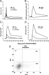
Flow cytometric analysis of CNS homing by systemic mononuclear cells. A–D, MOG-specific Th1, Th17, and Th17/Th1 (CD4+ IFN-γ+ IL-17+) cells in pooled spleen/lymph nodes (A), CNS (pooled brain and spinal cord) immune mononuclear cells (B), CNS Th1, Th17 and Th17/Th1 cells (C), and CNS Teff (Th1, Th17 and Th17/Th1) and Tregs (D). Data shown in A–D are means ± SEMs of at least three independent experiments at each time point. E, Example of the flow cytometric analysis of CNS CD4-gated mononuclear cells prepared from a day 14 post-CFA/MOG peptide mouse for intracellular expression of IFN-γ and IL-17, showing 3.4% Th1 cells (IFN-γ high, IL-17 low), 13.64% Th17 cells (IFN-γ low, IL-17 high) and 1.86% Th17/Th1 cells (IFN-γ high, IL-17 high).
Figure 3.
Figure 3.
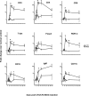
qRT/PCR for immune marker mRNAs in MOG peptide and CFA control spinal cords harvested on days −1 (normal control), 3, 7, 10, 14, 21, 35, and 98. The x axis shows days post-CFA/MOG (circles/solid line) or CFA (squares/dotted line). The y axis shows the ratio of abundance of each mRNA in MOG or CFA to that in normal controls. Fold changes are provided for T-cell surface markers (CD3, CD4, CD8), transcription factors associated with Th1 (T-bet), Th17 (ROR-γt), or Treg (Foxp3) T cells; and markers for B cells (IgM and CD19) and macrophages/microglia (CD11b). Data are means ± SEMs of at least three independent experiments. *Results in CFA/MOG peptide spinal cords were significantly higher than in CFA control spinal cords (p < 0.05); θ, results in CFA control spinal cords were significantly higher than in CFA/MOG peptide spinal cords (p < 0.05, Mann–Whitney U test).
Figure 4.
Figure 4.
Early perivascular and subpial neutrophil- and lymphocyte-rich inflammatory infiltrates in MOG peptide EAE mice. A, Expression of immunoreactive granulocyte colony-stimulating factor (G-CSF) on the spinal meninges on day 10 post-CFA/MOG peptide; this was no longer evident by day 12 post-CFA/MOG peptide, when a subpial infiltrate of Ly6G+ neutrophils had formed. B, Perivascular infiltrate of Ly6G+ neutrophils and CD3ε+ lymphocytes on day 12 post-CFA/MOG peptide. C, Another perivascular inflammatory infiltrate, containing Ly6G+/MMP8+ neutrophils, but without substantial recruitment to the infiltrate of IBA1+ microglia or monocyte-derived macrophages.
Figure 5.
Figure 5.
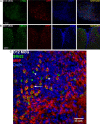
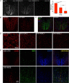
Axons are damaged within spinal cord inflammatory infiltrates in MOG peptide EAE mice. A, Ly6G+ neutrophil-rich inflammatory infiltrate containing APP+ damaged axons, from a day 14 post-CFA/MOG peptide mouse. B, By day 21 post-CFA/MOG peptide, SMI32+ and APP+ damaged axons are seen outside zones of overt infiltration by DAPI+ inflammatory cells; note the low overlap between SMI32 and APP immunoreactivities in this field. C, This field was in the ventrolateral lumbar spinal cord of a mouse given CFA/MOG peptide 12 d previously. Within a zone of DAPI nuclear-stained inflammatory cells, an SMI32+ axon still encircled by myelin basic protein (MBP) immunoreactivity is indicated by the arrow, a demyelinated SMI32+ axon by an arrowhead, and an apparently vacuolated SMI32+ axon surrounded by a ring of MBP immunoreactivity by an asterisk.
Figure 6.
Figure 6.
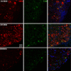
Accumulations of the endovesicular Toll-like receptor TLR8 in SMI32+ axons. Left, SMI32+ axons in ventrolateral spinal cord at days 12, 35, and 98 post-CFA/MOG peptide, respectively. Center, Same fields immunostained for TLR8. Right, Merged panels, with DAPI nuclear staining as well. Note that the ring-like pattern of SMI32 immunostaining is more prominent on day 12 post-CFA/MOG peptide, and that the SMI32 mAb and the anti-TLR8 antibody often bind to the same axons.
Figure 7.
Figure 7.
SMI32+ axons in MOG peptide EAE mice contain endolysosome-associated Rab7 and express inflammasome protein NALP1, which contributes to the processing of IL-1β. A, Ventrolateral spinal cord of a day 14 post-CFA/MOG peptide mouse immunostained (from left to right) for Rab7, SMI32, and the two markers together. The white arrowhead indicates an SMI32+ axon containing Rab7. B, Ventrolateral spinal cord of a day 21 post-CFA/MOG peptide mouse immunostained (from left to right) for NALP1, SMI32, and both markers. By 21 d post-CFA/MOG peptide, many SMI32+ axons expressed NALP1. C, Western blot demonstrating induction of IL-1β precursor (pro-IL-1β) and IL-1 β in spinal cords of day 14 post-CFA/MOG peptide mice. The asterisk indicates a nonspecific band.
Figure 8.
Figure 8.
Progressive loss of EYFP+ dorsal corticospinal axons in MOG peptide EAE. A, Optical sections (0.13 mm) of the L3 dorsal corticospinal tracts (dCSTs) of day 101 CFA control; day 35 CFA/MOG peptide; and day 101 CFA/MOG peptide EmxCre/Rosa-STOP-EYFP transgenic mice. The far right panel shows the total (right plus left) EYFP+ axon counts in the combined right plus left dorsal dCSTs at L3 of four normal or CFA controls; six d 35 CFA/MOG peptide mice; and four d 101 CFA/MOG peptide mice (means ± SDs). B, dCSTs of a day 101 post-CFA control and a day 101 post-CFA/MOG peptide EAE mouse, immunostained with SMI312, which recognizes multiple phosphorylated neurofilament epitopes; note the severe depletion of SMI312+ axons in the EAE specimen and that dCST axons are shown in green. C, dCSTs of day 35 post-CFA control and day 35 post-CFA/MOG peptide EAE EmxCre/Rosa-STOP-EYFP mice immunostained for the microglial/macrophage marker Iba1. Note the prominence of Iba1+ cells in the EAE specimen. D, SMI312 immunostained axons within the dCSTs of two day 101 post-CFA control mice (on the left) and two day 101 post-CFA/MOG peptide mice (on the right) in 0.13 mm optical sections, confirming severe dCST axonal depletion in chronic MOG peptide EAE. E, Greater prominence of both Iba1+ and CD68+ microglial/monocyte-derived macrophages in day 35 post-CFA/MOG peptide than day 35 post-CFA control EmxCre/Rosa-STOP-EYFP lumbar spinal cord dorsal funiculi. EYFP+ dCST axons are pseudocolored blue in this panel.

Similar articles

Cited by

References

    1. Aboul-Enein F, Weiser P, Höftberger R, Lassmann H, Bradl M. Transient axonal injury in the absence of demyelination: a correlate of clinical disease in acute experimental autoimmune encephalomyelitis. Acta Neuropathol. 2006;111:539–547. - PubMed
    1. Ahmed Z, Doward AI, Pryce G, Taylor DL, Pocock JM, Leonard JP, Baker D, Cuzner ML. A role for caspase-1 and -3 in the pathology of experimental allergic encephalomyelitis: inflammation versus degeneration. Am J Pathol. 2002;161:1577–1586. - PMC - PubMed
    1. Alelú-Paz R, Iturrieta-Zuazo I, Byne W, Haroutunian V, García-Villanueva M, Rábano A, García-Amado M, Prensa L, Giménez-Amaya JM. A new antigen retrieval technique for human brain tissue. PLoS One. 2008;3:e3378. - PMC - PubMed
    1. Amadio S, Pluchino S, Brini E, Morana P, Guerriero R, Boneschi FM, Comi G, Zaratin P, Muzio V, del Carro U. Motor evoked potentials in a mouse model of chronic multiple sclerosis. Muscle Nerve. 2006;33:265–273. - PubMed
    1. Annunziato F, Cosmi L, Santarlasci V, Maggi L, Liotta F, Mazzinghi B, Parente E, Filì L, Ferri S, Frosali F, Giudici F, Romagnani P, Parronchi P, Tonelli F, Maggi E, Romagnani S. Phenotypic and functional features of human Th17 cells. J Exp Med. 2007;204:1849–1861. - PMC - PubMed

Publication types

MeSH terms

Substances