Glutamine metabolism is essential for human cytomegalovirus infection
- PMID: 19939921
- PMCID: PMC2812398
- DOI: 10.1128/JVI.02123-09
Glutamine metabolism is essential for human cytomegalovirus infection
Abstract
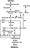
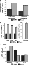
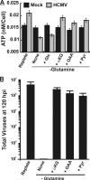
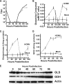
Human fibroblasts infected with human cytomegalovirus (HCMV) were more viable than uninfected cells during glucose starvation, suggesting that an alternate carbon source was used. We have determined that infected cells require glutamine for ATP production, whereas uninfected cells do not. This suggested that during infection, glutamine is used to fill the tricarboxylic acid (TCA) cycle (anaplerosis). In agreement with this, levels of glutamine uptake and ammonia production increased in infected cells, as did the activities of glutaminase and glutamate dehydrogenase, the enzymes needed to convert glutamine to alpha-ketoglutarate to enter the TCA cycle. Infected cells starved for glutamine beginning 24 h postinfection failed to produce infectious virions. Both ATP and viral production could be rescued in glutamine-starved cells by the TCA intermediates alpha-ketoglutarate, oxaloacetate, and pyruvate, confirming that in infected cells, a program allowing glutamine to be used anaplerotically is induced. Thus, HCMV infection activates the mechanisms needed to switch the anaplerotic substrate from glucose to glutamine to accommodate the biosynthetic and energetic needs of the viral infection and to allow glucose to be used biosynthetically.
Figures


Similar articles
-
Viral effects on metabolism: changes in glucose and glutamine utilization during human cytomegalovirus infection.Trends Microbiol. 2011 Jul;19(7):360-7. doi: 10.1016/j.tim.2011.04.002. Epub 2011 May 12. Trends Microbiol. 2011. PMID: 21570293 Free PMC article. Review.
-
Nitric Oxide Circumvents Virus-Mediated Metabolic Regulation during Human Cytomegalovirus Infection.mBio. 2020 Dec 15;11(6):e02630-20. doi: 10.1128/mBio.02630-20. mBio. 2020. PMID: 33323506 Free PMC article.
-
Viruses and metabolism: alterations of glucose and glutamine metabolism mediated by human cytomegalovirus.Adv Virus Res. 2011;80:49-67. doi: 10.1016/B978-0-12-385987-7.00003-8. Adv Virus Res. 2011. PMID: 21762821
-
Adaptation of renal tricarboxylic acid cycle metabolism to various acid-base states: study with [3-13C,5-15N]glutamine.Miner Electrolyte Metab. 1991;17(1):21-31. Miner Electrolyte Metab. 1991. PMID: 1770913
-
Carboxylation and anaplerosis in neurons and glia.Mol Neurobiol. 2000 Aug-Dec;22(1-3):21-40. doi: 10.1385/MN:22:1-3:021. Mol Neurobiol. 2000. PMID: 11414279 Review.
Cited by
-
Targeted Metabolic Reprogramming to Improve the Efficacy of Oncolytic Virus Therapy.Mol Ther. 2020 Jun 3;28(6):1417-1421. doi: 10.1016/j.ymthe.2020.03.014. Epub 2020 Mar 20. Mol Ther. 2020. PMID: 32243836 Free PMC article. Review.
-
HIV-1 pathogenicity and virion production are dependent on the metabolic phenotype of activated CD4+ T cells.Retrovirology. 2014 Nov 25;11:98. doi: 10.1186/s12977-014-0098-4. Retrovirology. 2014. PMID: 25421745 Free PMC article.
-
Preventing Allograft Rejection by Targeting Immune Metabolism.Cell Rep. 2015 Oct 27;13(4):760-770. doi: 10.1016/j.celrep.2015.09.036. Epub 2015 Oct 17. Cell Rep. 2015. PMID: 26489460 Free PMC article.
-
1H Nuclear Magnetic Resonance Metabolomics of Plasma Unveils Liver Dysfunction in Dengue Patients.J Virol. 2016 Jul 27;90(16):7429-7443. doi: 10.1128/JVI.00187-16. Print 2016 Aug 15. J Virol. 2016. PMID: 27279613 Free PMC article.
-
Glutamine Metabolism in Both the Oxidative and Reductive Directions Is Triggered in Shrimp Immune Cells (Hemocytes) at the WSSV Genome Replication Stage to Benefit Virus Replication.Front Immunol. 2019 Sep 4;10:2102. doi: 10.3389/fimmu.2019.02102. eCollection 2019. Front Immunol. 2019. PMID: 31555294 Free PMC article.
References
-
- Baggetto, L. G. 1992. Deviant energetic metabolism of glycolytic cancer cells. Biochimie 74:959-974. - PubMed
-
- Bergmeyer, H. U. 1984. Methods of enzymatic analysis, vol. III and IV. Verlag Chemie, Weinheim, Germany.
-
- Chakravarty, K., H. Cassuto, L. Reshef, and R. W. Hanson. 2005. Factors that control the tissue-specific transcription of the gene for phosphoenolpyruvate carboxykinase-C. Crit. Rev. Biochem. Mol. Biol. 40:129-154. - PubMed
-
- Cooper, E. H., P. Barkhan, and A. J. Hale. 1963. Observations on the proliferation of human leucocytes cultured with phytohaemagglutinin. Br. J. Haematol. 9:101-111. - PubMed
Publication types
MeSH terms
Substances
Grants and funding
LinkOut - more resources
Full Text Sources
Other Literature Sources
Medical

