Experimental diabetes mellitus exacerbates tau pathology in a transgenic mouse model of Alzheimer's disease
- PMID: 19936237
- PMCID: PMC2775636
- DOI: 10.1371/journal.pone.0007917
Experimental diabetes mellitus exacerbates tau pathology in a transgenic mouse model of Alzheimer's disease
Abstract
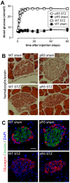
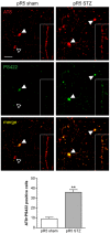
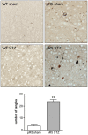
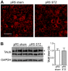
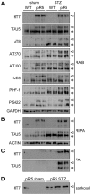
Diabetes mellitus (DM) is characterized by hyperglycemia caused by a lack of insulin, insulin resistance, or both. There is increasing evidence that insulin also plays a role in Alzheimer's disease (AD) as it is involved in the metabolism of beta-amyloid (Abeta) and tau, two proteins that form Abeta plaques and neurofibrillary tangles (NFTs), respectively, the hallmark lesions in AD. Here, we examined the effects of experimental DM on a pre-existing tau pathology in the pR5 transgenic mouse strain that is characterized by NFTs. pR5 mice express P301L mutant human tau that is associated with dementia. Experimental DM was induced by administration of streptozotocin (STZ), which causes insulin deficiency. We determined phosphorylation of tau, using immunohistochemistry and Western blotting. Solubility of tau was determined upon extraction with sarkosyl and formic acid, and Gallyas silver staining was employed to reveal NFTs. Insulin depletion by STZ administration in six months-old non-transgenic mice causes increased tau phosphorylation, without its deposition or NFT formation. In contrast, in pR5 mice this results in massive deposition of hyperphosphorylated, insoluble tau. Furthermore, they develop a pronounced tau-histopathology, including NFTs at this early age, while the pathology in sham-treated pR5 mice is moderate. Whereas experimental DM did not result in deposition of hyperphosphorylated tau in non-transgenic mice, a predisposition to develop a tau pathology in young pR5 mice was both sufficient and necessary to exacerbate tau deposition and NFT formation. Hence, DM can accelerate onset and increase severity of disease in individuals with a predisposition to developing tau pathology.
Conflict of interest statement
Figures
Similar articles
-
Sodium selenate mitigates tau pathology, neurodegeneration, and functional deficits in Alzheimer's disease models.Proc Natl Acad Sci U S A. 2010 Aug 3;107(31):13888-93. doi: 10.1073/pnas.1009038107. Epub 2010 Jul 19. Proc Natl Acad Sci U S A. 2010. PMID: 20643941 Free PMC article.
-
Divergent phosphorylation pattern of tau in P301L tau transgenic mice.Eur J Neurosci. 2008 Jul;28(1):137-47. doi: 10.1111/j.1460-9568.2008.06318.x. Eur J Neurosci. 2008. PMID: 18662339
-
Long term high fat diet induces metabolic disorders and aggravates behavioral disorders and cognitive deficits in MAPT P301L transgenic mice.Metab Brain Dis. 2022 Aug;37(6):1941-1957. doi: 10.1007/s11011-022-01029-x. Epub 2022 Jun 15. Metab Brain Dis. 2022. PMID: 35704147
-
Amyloid-induced neurofibrillary tangle formation in Alzheimer's disease: insight from transgenic mouse and tissue-culture models.Int J Dev Neurosci. 2004 Nov;22(7):453-65. doi: 10.1016/j.ijdevneu.2004.07.013. Int J Dev Neurosci. 2004. PMID: 15465275 Review.
-
Mouse models of Alzheimer's disease: the long and filamentous road.Neurol Res. 2003 Sep;25(6):590-600. doi: 10.1179/016164103101202020. Neurol Res. 2003. PMID: 14503012 Review.
Cited by
-
Altered regulation of Akt signaling with murine cerebral malaria, effects on long-term neuro-cognitive function, restoration with lithium treatment.PLoS One. 2012;7(10):e44117. doi: 10.1371/journal.pone.0044117. Epub 2012 Oct 17. PLoS One. 2012. PMID: 23082110 Free PMC article.
-
Benfotiamine prevents increased β-amyloid production in HEK cells induced by high glucose.Neurosci Bull. 2012 Oct;28(5):561-6. doi: 10.1007/s12264-012-1264-0. Epub 2012 Sep 8. Neurosci Bull. 2012. PMID: 22961478 Free PMC article.
-
The Angiotensin II Type 2 Receptor in Brain Functions: An Update.Int J Hypertens. 2012;2012:351758. doi: 10.1155/2012/351758. Epub 2012 Dec 25. Int J Hypertens. 2012. PMID: 23320146 Free PMC article.
-
ENU mutagenesis screen to establish motor phenotypes in wild-type mice and modifiers of a pre-existing motor phenotype in tau mutant mice.J Biomed Biotechnol. 2011;2011:130947. doi: 10.1155/2011/130947. Epub 2011 Dec 15. J Biomed Biotechnol. 2011. PMID: 22219655 Free PMC article. Review.
-
Neuronal models for studying tau pathology.Int J Alzheimers Dis. 2010 Jul 19;2010:528474. doi: 10.4061/2010/528474. Int J Alzheimers Dis. 2010. PMID: 20721342 Free PMC article.
References
-
- Gotz J, Streffer JR, David D, Schild A, Hoerndli F, et al. Transgenic animal models of Alzheimer's disease and related disorders: histopathology, behavior and therapy. Mol Psychiatry. 2004;9:664–683. - PubMed
-
- Ballatore C, Lee VM, Trojanowski JQ. Tau-mediated neurodegeneration in Alzheimer's disease and related disorders. Nat Rev Neurosci. 2007;8:663–672. - PubMed
-
- Lee VM, Goedert M, Trojanowski JQ. Neurodegenerative tauopathies. Annu Rev Neurosci. 2001;24:1121–1159. - PubMed
-
- Selkoe DJ. Alzheimer's disease: genotypes, phenotypes, and treatments. Science. 1997;275:630–631. - PubMed
Publication types
MeSH terms
Substances
LinkOut - more resources
Full Text Sources
Medical
Molecular Biology Databases

