

Cell fate decisions are specified by the dynamic ERK interactome
- PMID: 19935650
- PMCID: PMC3839079
- DOI: 10.1038/ncb1994
Cell fate decisions are specified by the dynamic ERK interactome
Erratum in
-
Author Correction: Cell fate decisions are specified by the dynamic ERK interactome.Nat Cell Biol. 2022 Mar;24(3):400. doi: 10.1038/s41556-022-00867-2. Nat Cell Biol. 2022. PMID: 35181749 No abstract available.
Abstract
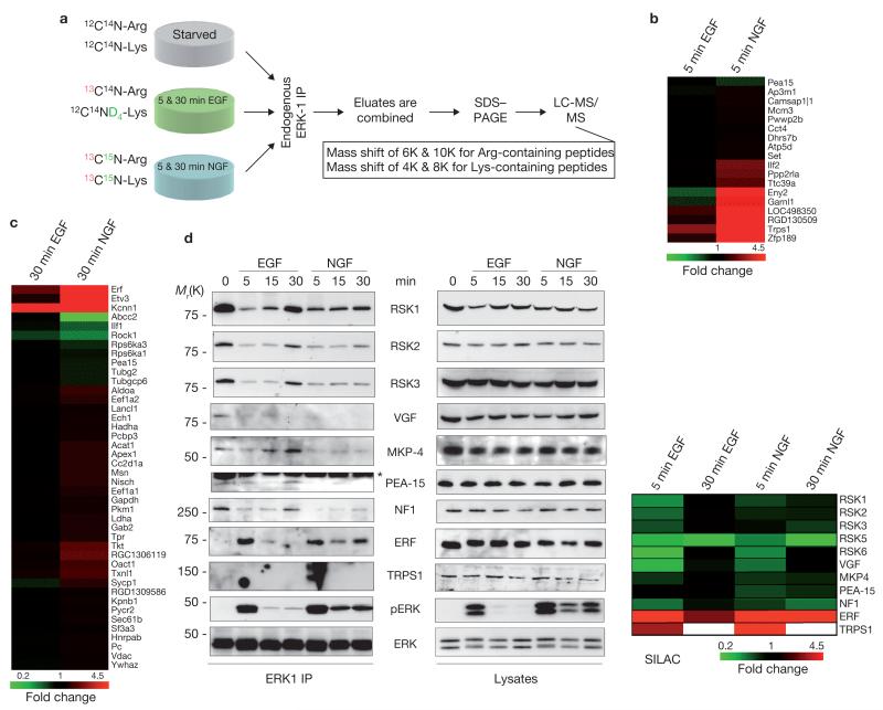
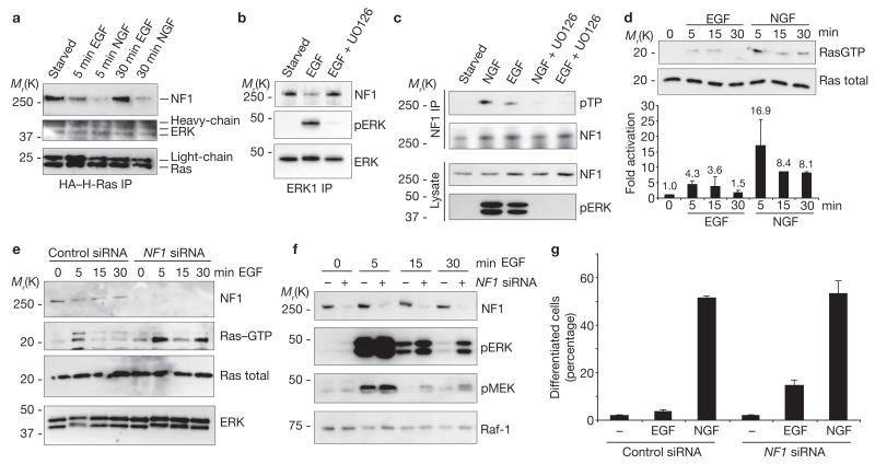
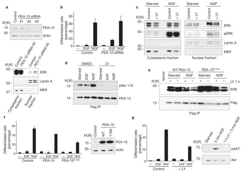
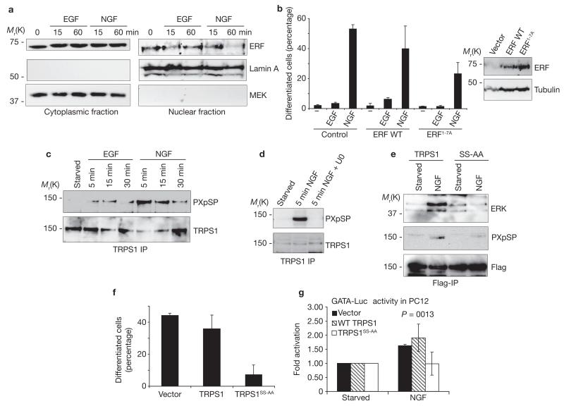
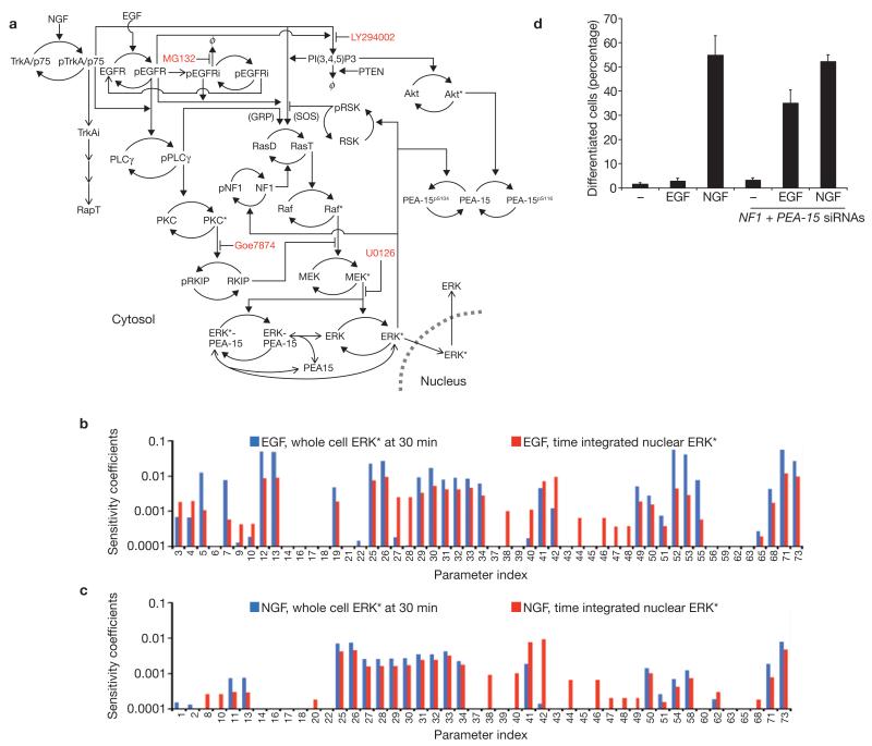
Extracellular signal-regulated kinase (ERK) controls fundamental cellular functions, including cell fate decisions. In PC12, cells shifting ERK activation from transient to sustained induces neuronal differentiation. As ERK associates with both regulators and effectors, we hypothesized that the mechanisms underlying the switch could be revealed by assessing the dynamic changes in ERK-interacting proteins that specifically occur under differentiation conditions. Using quantitative proteomics, we identified 284 ERK-interacting proteins. Upon induction of differentiation, 60 proteins changed their binding to ERK, including many proteins that were not known to participate in differentiation. We functionally characterized a subset, showing that they regulate the pathway at several levels and by different mechanisms, including signal duration, ERK localization, feedback, crosstalk with the Akt pathway and differential interaction and phosphorylation of transcription factors. Integrating these data with a mathematical model confirmed that ERK dynamics and differentiation are regulated by distributed control mechanisms rather than by a single master switch.
Figures
Similar articles
-
Sustained activation of M-Ras induced by nerve growth factor is essential for neuronal differentiation of PC12 cells.Genes Cells. 2006 Sep;11(9):1097-113. doi: 10.1111/j.1365-2443.2006.01002.x. Genes Cells. 2006. PMID: 16923128
-
Kinetics of receptor tyrosine kinase activation define ERK signaling dynamics.Sci Signal. 2020 Aug 18;13(645):eaaz5267. doi: 10.1126/scisignal.aaz5267. Sci Signal. 2020. PMID: 32817373 Free PMC article.
-
Sustained activation of extracellular signal-regulated kinase by nerve growth factor regulates c-fos protein stabilization and transactivation in PC12 cells.J Neurochem. 2006 Dec;99(6):1480-93. doi: 10.1111/j.1471-4159.2006.04250.x. J Neurochem. 2006. PMID: 17223854
-
Dissecting Cell-Fate Determination Through Integrated Mathematical Modeling of the ERK/MAPK Signaling Pathway.Methods Mol Biol. 2017;1487:409-432. doi: 10.1007/978-1-4939-6424-6_29. Methods Mol Biol. 2017. PMID: 27924583 Review.
-
The regulation of extracellular signal-regulated kinase (ERK) in mammalian cells.Int J Biochem Cell Biol. 2008;40(12):2707-19. doi: 10.1016/j.biocel.2008.04.009. Epub 2008 May 15. Int J Biochem Cell Biol. 2008. PMID: 18562239 Review.
Cited by
-
Microphthalmia-associated transcription factor in melanoma development and MAP-kinase pathway targeted therapy.Pigment Cell Melanoma Res. 2015 Jul;28(4):390-406. doi: 10.1111/pcmr.12370. Epub 2015 Apr 17. Pigment Cell Melanoma Res. 2015. PMID: 25818589 Free PMC article. Review.
-
Kinetics of gene derepression by ERK signaling.Proc Natl Acad Sci U S A. 2013 Jun 18;110(25):10330-5. doi: 10.1073/pnas.1303635110. Epub 2013 Jun 3. Proc Natl Acad Sci U S A. 2013. PMID: 23733957 Free PMC article.
-
Loss of FBXO31-mediated degradation of DUSP6 dysregulates ERK and PI3K-AKT signaling and promotes prostate tumorigenesis.Cell Rep. 2021 Oct 19;37(3):109870. doi: 10.1016/j.celrep.2021.109870. Cell Rep. 2021. PMID: 34686346 Free PMC article.
-
Mathematical model of a telomerase transcriptional regulatory network developed by cell-based screening: analysis of inhibitor effects and telomerase expression mechanisms.PLoS Comput Biol. 2014 Feb 13;10(2):e1003448. doi: 10.1371/journal.pcbi.1003448. eCollection 2014 Feb. PLoS Comput Biol. 2014. PMID: 24550717 Free PMC article.
-
Signaling Pathways in Proton and Non-proton ASIC1a Activation.Front Cell Neurosci. 2021 Oct 5;15:735414. doi: 10.3389/fncel.2021.735414. eCollection 2021. Front Cell Neurosci. 2021. PMID: 34675777 Free PMC article.
References
-
- Shaul YD, Seger R. The MEK/ERK cascade: from signaling specificity to diverse functions. Biochim. Biophys. Acta. 2007;1773:1213–1226. - PubMed
-
- Galabova-Kovacs G, et al. ERK and beyond: insights from B-Raf and Raf-1 conditional knockouts. Cell Cycle. 2006;5:1514–1518. - PubMed
-
- Marshall CJ. Specificity of receptor tyrosine kinase signaling: transient versus sustained extracellular signal-regulated kinase activation. Cell. 1995;80:179–185. - PubMed
-
- Yoon S, Seger R. The extracellular signal-regulated kinase: multiple substrates regulate diverse cellular functions. Growth Factors. 2006;24:21–44. - PubMed
Publication types
MeSH terms
Substances
Grants and funding
- 097945/WT_/Wellcome Trust/United Kingdom
- G0301131/MRC_/Medical Research Council/United Kingdom
- 073980/WT_/Wellcome Trust/United Kingdom
- 083524/WT_/Wellcome Trust/United Kingdom
- 081361/WT_/Wellcome Trust/United Kingdom
- G0801738/MRC_/Medical Research Council/United Kingdom
- BB/C511613/1/BB_/Biotechnology and Biological Sciences Research Council/United Kingdom
- WT_/Wellcome Trust/United Kingdom
- BB/C511572/1/BB_/Biotechnology and Biological Sciences Research Council/United Kingdom
- CRUK_/Cancer Research UK/United Kingdom
- 037538/WT_/Wellcome Trust/United Kingdom
- C12944/BB_/Biotechnology and Biological Sciences Research Council/United Kingdom
- C08577/BB_/Biotechnology and Biological Sciences Research Council/United Kingdom
LinkOut - more resources
Full Text Sources
Other Literature Sources
Molecular Biology Databases
Research Materials
Miscellaneous
