

Upregulation of immunoglobulin-related genes in cortical sections from multiple sclerosis patients
- PMID: 19919606
- PMCID: PMC8094770
- DOI: 10.1111/j.1750-3639.2009.00343.x
Upregulation of immunoglobulin-related genes in cortical sections from multiple sclerosis patients
Abstract
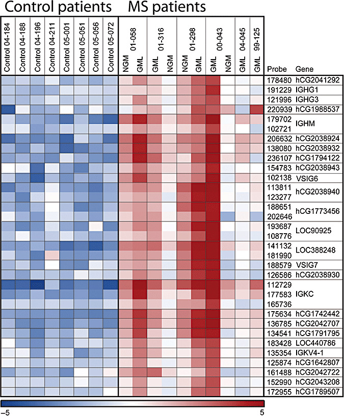
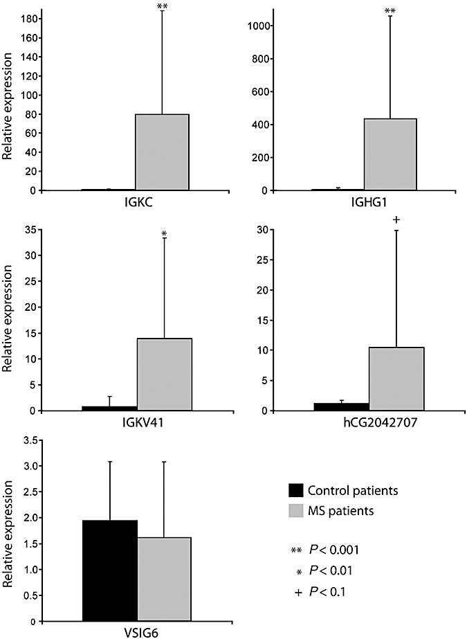
Multiple sclerosis (MS) is a demyelinating disease of the central nervous system (CNS). Microarray-based global gene expression profiling is a promising method, used to study potential genes involved in the pathogenesis of the disease. In the present study, we have examined global gene expression in normal-appearing gray matter and gray matter lesions from the cortex of MS patients, and compared them with cortical gray matter samples from controls. We observed a massive upregulation of immunoglobulin (Ig)-related genes in cortical sections of MS patients. Using immunohistochemistry, the activation of Ig genes seems to occur within plasma cells in the meninges. As synthesis of oligoclonal IgGs has been hypothesized to be caused by the activation of Epstein-Barr virus (EBV)-infected B-cells, we screened the brain samples for the presence of EBV by real-time quantitative polymerase chain reaction (qPCR) and immunohistochemistry, but no evidence of active or latent EBV infection was detected. This study demonstrates that genes involved in the synthesis of Igs are upregulated in MS patients and that this activation is caused by a small number of meningeal plasma cells that are not infected by EBV. The findings indicate that the Ig-producing B-cells found in the cerebrospinal fluid (CSF) of MS patients could have meningeal origin.
Figures


Similar articles
-
Meningeal inflammation is not associated with cortical demyelination in chronic multiple sclerosis.J Neuropathol Exp Neurol. 2009 Sep;68(9):1021-8. doi: 10.1097/NEN.0b013e3181b4bf8f. J Neuropathol Exp Neurol. 2009. PMID: 19680141
-
B-cell enrichment and Epstein-Barr virus infection in inflammatory cortical lesions in secondary progressive multiple sclerosis.J Neuropathol Exp Neurol. 2013 Jan;72(1):29-41. doi: 10.1097/NEN.0b013e31827bfc62. J Neuropathol Exp Neurol. 2013. PMID: 23242282
-
Transcriptional profile and Epstein-Barr virus infection status of laser-cut immune infiltrates from the brain of patients with progressive multiple sclerosis.J Neuroinflammation. 2018 Jan 16;15(1):18. doi: 10.1186/s12974-017-1049-5. J Neuroinflammation. 2018. PMID: 29338732 Free PMC article.
-
The histopathology of grey matter demyelination in multiple sclerosis.Acta Neurol Scand Suppl. 2009;(189):51-7. doi: 10.1111/j.1600-0404.2009.01216.x. Acta Neurol Scand Suppl. 2009. PMID: 19566500 Review.
-
Low intrathecal antibody production despite high seroprevalence of Epstein-Barr virus in multiple sclerosis: a review of the literature.J Neurol. 2018 Feb;265(2):239-252. doi: 10.1007/s00415-017-8656-z. Epub 2017 Nov 2. J Neurol. 2018. PMID: 29098417 Review.
Cited by
-
Common transcriptional signatures in brain tissue from patients with HIV-associated neurocognitive disorders, Alzheimer's disease, and Multiple Sclerosis.J Neuroimmune Pharmacol. 2012 Dec;7(4):914-26. doi: 10.1007/s11481-012-9409-5. Epub 2012 Oct 12. J Neuroimmune Pharmacol. 2012. PMID: 23065460 Free PMC article. Review.
-
A Review of Compartmentalised Inflammation and Tertiary Lymphoid Structures in the Pathophysiology of Multiple Sclerosis.Biomedicines. 2022 Oct 17;10(10):2604. doi: 10.3390/biomedicines10102604. Biomedicines. 2022. PMID: 36289863 Free PMC article. Review.
-
Contribution of vitamin D insufficiency to the pathogenesis of multiple sclerosis.Ther Adv Neurol Disord. 2013 Mar;6(2):81-116. doi: 10.1177/1756285612473513. Ther Adv Neurol Disord. 2013. PMID: 23483715 Free PMC article.
-
Trigger, pathogen, or bystander: the complex nexus linking Epstein- Barr virus and multiple sclerosis.Mult Scler. 2012 Sep;18(9):1204-8. doi: 10.1177/1352458512448109. Epub 2012 Jun 8. Mult Scler. 2012. PMID: 22685062 Free PMC article. Review.
-
Peli1 promotes microglia-mediated CNS inflammation by regulating Traf3 degradation.Nat Med. 2013 May;19(5):595-602. doi: 10.1038/nm.3111. Epub 2013 Apr 21. Nat Med. 2013. PMID: 23603814 Free PMC article.
References
-
- Bo L, Geurts JJG, Ravid R, Barkhof F (2004) Magnetic resonance imaging as a tool to examine the neuropathology of multiple sclerosis. Neuropathol Appl Neurobiol 30:106–117. - PubMed
-
- Bo L, Vedeler CA, Nyland H, Trapp BD, Mork SJ (2003) Intracortical multiple sclerosis lesions are not associated with increased lymphocyte infiltration. Mult Scler 9:323–331. - PubMed
-
- Bo L, Vedeler CA, Nyland HI, Trapp BD, Mork SJ (2003) Subpial demyelination in the cerebral cortex of multiple sclerosis patients. J Neuropathol Exp Neurol 62:723–732. - PubMed
-
- Breij EC, Brink BP, Veerhuis R, Van Den Berg C, Vloet R, Yan R et al (2008) Homogeneity of active demyelinating lesions in established multiple sclerosis. Ann Neurol 63:16–25. - PubMed
Publication types
MeSH terms
LinkOut - more resources
Full Text Sources
Medical
Research Materials
