. 2010 Jan 8;106(1):102-10.
doi: 10.1161/CIRCRESAHA.109.210914. Epub 2009 Nov 12.

Cardioprotection by CaMKII-deltaB is mediated by phosphorylation of heat shock factor 1 and subsequent expression of inducible heat shock protein 70

Affiliations

Cardioprotection by CaMKII-deltaB is mediated by phosphorylation of heat shock factor 1 and subsequent expression of inducible heat shock protein 70

Wei Peng et al. Circ Res. .

Abstract

Rationale: Ca2+/calmodulin-dependent protein kinase (CaMK)II is a multifunctional kinase involved in vital cellular processes such as Ca(2+) handling and cell fate regulation. In mammalian heart, 2 primary CaMKII isoforms, deltaB and deltaC, localize in nuclear and cytosolic compartments, respectively. Although previous studies have established an essential role of CaMKII-deltaC in cardiomyocyte apoptosis, the functional role of the more abundant isoform, CaMKII-deltaB, remains elusive.

Objective: Here, we determined the potential role of CaMKII-deltaB in regulating cardiomyocyte viability and explored the underlying mechanism.

Methods and results: In cultured neonatal rat cardiomyocytes, the expression of CaMKII-deltaB and CaMKII-deltaC was inversely regulated in response to H2O2-induced oxidative stress with a profound reduction of the former and an increase of the later. Similarly, in vivo ischemia/reperfusion (IR) led to an opposite regulation of these CaMKII isoforms in a rat myocardial IR model. Notably, overexpression of CaMKII-deltaB protected cardiomyocytes against oxidative stress-, hypoxia-, and angiotensin II-induced apoptosis, whereas overexpression of its cytosolic counterpart promoted apoptosis. Using cDNA microarray, real-time PCR and Western blotting, we demonstrated that overexpression of CaMKII-deltaB but not CaMKII-deltaC elevated expression of heat shock protein (HSP)70 family members, including inducible (i)HSP70 and its homolog (Hst70). Moreover, overexpression of CaMKII-deltaB led to phosphorylation and activation of heat shock factor (HSF)1, the primary transcription factor responsible for HSP70 gene regulation. Importantly, gene silencing of iHSP70, but not Hst70, abolished CaMKII-deltaB-mediated protective effect, indicating that only iHSP70 was required for CaMKII-deltaB elicited antiapoptotic signaling.

Conclusions: We conclude that cardiac CaMKII-deltaB and CaMKII-deltaC were inversely regulated in response to oxidative stress and IR injury, and that in contrast to CaMKII-deltaC, CaMKII-deltaB serves as a potent suppressor of cardiomyocyte apoptosis triggered by multiple death-inducing stimuli via phosphorylation of HSF1 and subsequent induction of iHSP70, marking both CaMKII-delta isoforms as promising therapeutic targets for the treatment of ischemic heart disease.


Figures
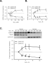
Figure 1
Figure 1. CaMKII-δB and CaMKII-δC expression in neonatal rat cardiomyocytes in response to oxidative stress or in rat myocardium subjected to ischemia/reperfusion (IR) injury
A. CaMKII-δB and CaMKII-δC gene expression assayed by real-time PCR in response to different concentrations of H2O2 treatment for 8 h in cultured neonatal cardiomyocytes (n = 6 for each data point). B. CaMKII-δB and CaMKII-δC gene expression assayed by real-time PCR in response to H2O2 (200 μM) treatment for different period of time in cultured neonatal cardiomyocytes (n = 6 for each data point). C. Typical Western blots and the statistic data of the time course of CaMKII-δ protein abundance in nuclear and cytosolic fractions from neonatal cardiomyocytes treated with H2O2 (200 μM) (n = 4 for each time point). D. shows CaMKII-δB and CaMKII-δC mRNA levels in ischemic area of rat hearts subjected to 45 min ischemia followed by different periods of reperfusion assayed by real-time PCR (n = 6 for each time point). E. CaMKII-δB and CaMKII-δC protein levels assayed by Western blotting in nuclear and cytosolic fractions from ischemic myocardium of rat hearts (n = 3-5 for each time point). For all panels, * P<0.05; † P<0.01. vs. baseline.
Figure 2
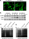
Figure 2. Overexpression of CaMKII-δB protects both neonatal and adult rat cardiomyocytes against H2O2-triggered apoptosis
A. Immunofluorescent imaging with an anti-HA antibody to visualize the intracellular distribution of HA-tagged CaMKII-δB and CaMKII-δC in neonatal rat cardiomyocytes infected with Adv-CaMKII-δB or Adv-CaMKII-δC (both at 20 m.o.i. for 24 h). B. Typical Western blot with a site-specific antibody reacting with CaMKII-d or phosphorylated CaMKII-d in lysate from myocytes infected with Adv-β-gal, Adv-CaMKII-δB, or Adv-CaMKII-δC at m.o.i. as indicated for 24 h. C. DNA laddering in neonatal rat cardiomyocytes infected with Adv-CaMKII-δB, Adv-CaMKII-δC or Adv-β-gal (all at 20 m.o.i. for 24 h) then treated with H2O2 (200 μM) for another 24 h. Similar results were obtained in three independent experiments. D. Representative DNA laddering in adult rat cardiomyocytes infected with Adv-CaMKII-δB or Adv-β-gal (both at 100 m.o.i. for 24 h) then treated with H2O2 (10 μM) for another 24 h. Similar results were obtained in three independent experiments. E. Average data of Hoechst staining in neonatal cardiomyocytes infected with Adv-CaMKII-δB, Adv-CaMKII-δC or Adv-β-gal (all at 20 m.o.i. for 24 h) then subjected to H2O2 (200 μM) for 24 h. F. Average data of Hoechst staining in neonatal cardiomyocytes infected with Adv-CaMKII-δB or Adv-β-gal (at 20 m.o.i. for 24 h) then subjected to hypoxia for 9 h. G. Average data of Hoechst staining in neonatal cardiomyocytes infected with Adv-CaMKII-δB or Adv-β-gal (at 20 m.o.i. for 24 h then subjected to Ang II (1 μM for 48 h). For E-G, each data point shows the result from 5000-8000 cells in four independent experiments for Hoechst staining (* P<0.01 vs. Adv-β-gal in the absence of H2O2; † P<0.01 vs. the corresponding Adv-β-gal in each group of different concentrations of H2O2 or as indicated).
Figure 3
Figure 3. Upregulation of iHSP70 and Hst70 by overexpression of CaMKII-δB but not CaMKII-δC in cultured neonatal rat cardiomyocytes
A. iHSP70 and Hst70 gene expression assayed by cDNA microarray analysis in cells infected with Adv-CaMKII-δB, Adv-CaMKII-δC, or Adv-β-gal. B. iHSP70 gene expression assayed by real-time PCR in neonatal cardiomyocytes after adenoviral infection for 36 h or 48 h. (n = 4). C. Typical Western blot with an anti-iHSP70 antibody and the average data in cells infected with Adv-CaMKII-δB, Adv-CaMKII-δC, or Adv-β-gal (at 20 m.o.i.) for 48 h. (n = 4). For all panels, * P<0.05 vs. Adv-CaMKII-δC and Adv-β-gal.
Figure 4
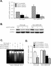
Figure 4. Gene silencing of iHSP70 blocks CaMKII-δB-mediated anti-apoptotic effect
A. siRNA-induced reduction of iHSP70 at mRNA level assayed by real-time PCR in neonatal cardiomyocytes infected with Adv-β-gal or Adv-CaMKII-δB (n = 3 for each group * P<0.05 vs. the NC of each group). B. siRNA-induced reduction of iHSP70 at protein level in cells infected with Adv-β-gal or Adv-CaMKII-δB. Similar results were observed in three independent experiments. C. DNA laddering in cells infected with Adv-β-gal or Adv-CaMKII-δB in the absence or presence of iHSP70 siRNA after H2O2 (200 μM for 24 h) treatment. Similar results were obtained in three independent experiments. D. MTT cell viability assay in cells infected with Adv-β-gal or Adv-CaMKII-δB in the absence or presence of iHSP70 siRNA after H2O2 (200 μM for 24 h) treatment (n = 3, * P<0.05 vs. the NC of each group).
Figure 5
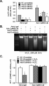
Figure 5. Gene silencing of Hst70 does not affect CaMKII-δB-mediated anti-apoptotic effect
A. siRNA-induced reduction of Hst70 at mRNA level assayed by real-time PCR in neonatal cardiomyocytes infected with Adv-β-gal or Adv-CaMKII-δB (n = 3 for each group, * P<0.05 vs. the NC of each group). B. DNA laddering in myocytes infected with Adv-β-gal or Adv-CaMKII-δB in the absence or presence of Hst70 siRNA after H2O2 (200 μM for 24 h) treatment. C. MTT cell viability assay in cells infected with Adv-β-gal or Adv-CaMKII-δB in the absence or presence of Hst70 siRNA after H2O2 (200 μM for 24 h) treatment (n = 3, * P<0.05 vs. the NC of each group).
Figure 6
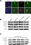
Figure 6. CaMKII-δB co-localizes with HSF1 and selectively increases phosphorylation of HSF1 at Ser230
A. Confocal immunofluorescent imaging to visualize the nucleus by DAPI staining (blue) and intracellular distribution of HSF1 (green) and HA-tagged CaMKII-δB (red) in cardiomyocytes infected with Adv-CaMKII-δB (scale bar is 20 μm). The merged imaging (yellow) shows HSF1 and HA-tagged CaMKII-δB were co-localized in the nuclear compartment. B. Typical western blots with an antibody reacting with total or Ser230- or Ser303-phosphorylated HSF1 in uninfected cells (Control) or those infected with Adv-CaMKII-δB, Adv-CaMKII-δC, or Adv-β-gal (at 20 m.o.i.). Similar results were obtained in other three independent experiments. C. Phosphorylation of HSF1 at Ser230 in neonatal cardiomyocytes infected with Adv-CaMKII-δB for 36 h and then treated with H2O2 (200 μM) for various time periods.
Figure 7
Figure 7. Schematic presentation to show oxidative stress- or IR-induced opposite regulation of the expression and functional consequence of the two major cardiac CaMKII isoforms, CaMKII-δB and CaMKII-δC
Oxidative stress or IR injury leads to a downregulation of the anti-apoptotic isoform, CaMKII-δB, and an upregulation of the apoptotic isoform, CaMKII-δC, resulting in robust cardiomyocyte apoptosis. The anti-apoptotic effect of CaMKII-δB is attributable to the kinase-mediated phosphorylation of HSF1 at Ser230 and subsequent induction of iHSP70 gene expression.

Similar articles

Cited by

References

    1. Edman CF, Schulman H. Identification and characterization of delta B-CaM kinase and delta C-CaM kinase from rat heart, two new multifunctional Ca2+/calmodulin-dependent protein kinase isoforms. Biochim Biophys Acta. 1994;1221:89–101. - PubMed
    1. Baltas LG, Karczewski P, Krause EG. The cardiac sarcoplasmic reticulum phospholamban kinase is a distinct delta-CaM kinase isozyme. FEBS Lett. 1995;373:71–75. - PubMed
    1. Hoch B, Meyer R, Hetzer R, Krause EG, Karczewski P. Identification and expression of delta-isoforms of the multifunctional Ca2+/calmodulin-dependent protein kinase in failing and nonfailing human myocardium. Circ Res. 1999;84:713–721. - PubMed
    1. Hoch B, Wobus AM, Krause EG, Karczewski P. delta-Ca(2+)/calmodulin-dependent protein kinase II expression pattern in adult mouse heart and cardiogenic differentiation of embryonic stem cells. J Cell Biochem. 2000;79:293–300. - PubMed
    1. Srinivasan M, Edman CF, Schulman H. Alternative splicing introduces a nuclear localization signal that targets multifunctional CaM kinase to the nucleus. J Cell Biol. 1994;126:839–852. - PMC - PubMed

Publication types

MeSH terms