. 2010 Mar;18(3):455-63.
doi: 10.1016/j.joca.2009.10.014. Epub 2009 Nov 4.

10mM glucosamine prevents activation of proADAMTS5 (aggrecanase-2) in transfected cells by interference with post-translational modification of furin

Affiliations

10mM glucosamine prevents activation of proADAMTS5 (aggrecanase-2) in transfected cells by interference with post-translational modification of furin

D R McCulloch et al. Osteoarthritis Cartilage. 2010 Mar.

Abstract

Objective: Glucosamine has been previously shown to suppress cartilage aggrecan catabolism in explant cultures. We determined the effect of glucosamine on ADAMTS5 (a disintegrin-like and metalloprotease domain (reprolysin type) with thrombospondin type-1 motifs 5), a major aggrecanase in osteoarthritis, and investigated a potential mechanism underlying the observed effects.

Design: HEK293F and CHO-K1 cells transiently transfected with ADAMTS5 cDNA were treated with glucosamine or the related hexosamine mannosamine. Glucosamine effects on FURIN transcription were determined by quantitative RT-PCR. Effects on furin-mediated processing of ADAMTS5 zymogen, and aggrecan processing by glucosamine-treated cells, were determined by western blotting. Post-translational modification of furin and N-glycan deficient furin mutants generated by site-directed mutagenesis was analyzed by western blotting, and the mutants were evaluated for their ADAMTS5 processing ability in furin-deficient CHO-RPE.40 cells.

Results: Ten mM glucosamine and 5-10mM mannosamine reduced excision of the ADAMTS5 propeptide, indicating interference with the propeptide excision mechanism, although mannosamine compromised cell viability at these doses. Although glucosamine had no effect on furin mRNA levels, western blot of furin from glucosamine-treated cells suggested altered post-translational modification. Glucosamine treatment led to decreased glycosylation of cellular furin, with reduced furin autoactivation as the consequence. Recombinant furin treated with peptide N-glycanase F had reduced activity against a synthetic peptide substrate. Indeed, site-directed mutagenesis of two furin N-glycosylation sites, Asn(387) and Asn(440), abrogated furin activation and this mutant was unable to rescue ADAMTS5 processing in furin-deficient cells.

Conclusions: Ten mM glucosamine reduces excision of the ADAMTS5 propeptide via interference with post-translational modification of furin and leads to reduced aggrecanase activity of ADAMTS5.


Conflict of interest statement

None of the authors have any conflict of interest relating to the submitted manuscript.

Figures

Figure 1
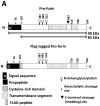
Figure 1. ADAMTS5 constructs used in the analysis of glucosamine effects
Domain organization of ADAMTS5 and ADAMTS5 ProCat, predicted molecular weights of the relevant protein species, N-linked glycosylation sites and furin processing sites are shown. The key to the various modules is at the bottom of the figure.
Figure 2
Figure 2. ADAMTS5 activation is inhibited by glucosamine treatment
A. Treatment of HEK293F cells transiently transfected with ADAMTS5 using 5 mM or 10 mM glucosamine results in increased uncleaved zymogen (top two panels) and decreased processed propeptide (Pro, second panel from the bottom). Western blotting with anti-GAPDH (bottom panel) was used to indicate sample loading. B. Quantitation of the effects of 10 mM glucosamine on the levels of ADAMTS5 zymogen and cleaved propeptide from 3 independent experiments shows a statistically significant effect of glucosamine on zymogen cleavage (*p<0.05; Student t-test, error bars represent S.E.M.). C. Treatment with 10 mM glucosamine results in loss of aggrecanase activity in the medium of ADAMTS5 transfected HEK293F cells. The top two panels illustrate reduced zymogen processing, whereas the anti-NITEGE373 immunoblot (second panel from the bottom) shows reduced aggrecanase activity in medium of glucosamine treated cells.
Figure 3
Figure 3. Effect of mannosamine on ADAMTS5 propeptide processing, and cell viability after glucosamine and mannosamine treatment with various doses
A. Treatment of ADAMTS5-transfected HEK293F cells with 5 mM, and 10 mM mannosamine resulted in a dose-dependent inhibition of ADAMTS5 zymogen processing. GAPDH western blot suggests decreased cell numbers with 5–10 mM mannosamine (bottom panel). B. Trypan blue exclusion assay shows that 5–10 mM mannosamine and 25 mM glucosamine, but not 10 mM glucosamine had a significant decrease in cell viability (the data are presented as the mean ± S.D. of ten cell counts from duplicate and control hexosamine-treated cultures).
Figure 4
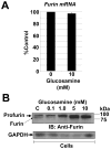
Figure 4. Glucosamine affects post-translational modification of furin
A. Furin mRNA levels in HEK293F cells treated with 10 mM glucosamine were similar to those of untreated cells. 18s ribosomal RNA levels were used for normalization of furin mRNA. The data are presented as the mean ± S.D. from three independent experiments. B. Treatment with 5mM and 10 mM glucosamine alters electrophoretic migration of furin in HEK293F cells. The major furin species seen with 5mM and 10 mM glucosamine treatments corresponds in size to active furin.
Figure 5
Figure 5. Deglycosylation of purified recombinant furin reduces its activity
A. Kinetic data for two independent assays are plotted as a function of fluorescence (AMC liberation) versus time at 25 °C. Each curve represents the mean kinetic data from triplicate reactions for each deglycosylated furin preparation (HI=heat-inactivated). B. Activity of glycosylated versus deglycosylated furin towards the fluorogenic substrate Boc-RVRR-AMC was measured at 10 minutes. The liberation of AMC by furin was significantly decreased by the removal of N-linked sugars with PNGaseF. C. Western blot of recombinant furin following treatment with PNGase F, showing complete deglycosylation.
Figure 6
Figure 6. Mutation of the N-glycosylation sites of furin inhibits its autoactivation
A. Schematic of human furin constructs used in the study depicting their domain structure (key to various domains is at the bottom), predicted molecular weights, autocatalytic and C-terminal cleavage sites and N-linked glycosylation sites. B. Detection of transiently expressed N-terminal FLAG-tagged WT-furin with or without dec-RVKR-CMK as well as FLAG-tagged Asn387Gln, Asn440Gln, Asn387+440Gln furin mutants in HEK293F cells using anti-FLAG M1 antibody. Note enhanced in-gel migration of the mutants, complete loss of activation of Asn387+440Gln furin, and reduced activation of Asn440Gln furin. Asn387+440Gln furin migrates as a species intermediate in size between wild-type profurin and furin. C. Shed furin was detected in the conditioned medium of the cells shown in B using a polyclonal anti-FLAG antibody, where similar relative migration of the various constructs is seen as in cells, albeit with reduction of molecular mass owing to C-terminal processing. The active form of furin is not detected using this antibody. D. Electrophoretic mobility of shed furin from transfected cells treated with or without 10 mM glucosamine was compared alongside the single and double N-glycosylation mutants, suggestive of impaired N-glycosylation of wild-type furin in the presence of glucosamine. E. Western blot analysis of the samples shown in panel D was done following enzymatic deglycosylation with PNGAse F. Note that the 10 mM glucosamine treated furin and glycosylation mutant furin co-migrated with deglycosylated wild-type (WT) profurin, as well as with profurin from RVKR treated cells expressing wild-type furin, indicating retention of the propeptide following 10 mM glucosamine treatment (compare with 6D). The identity of the 90 kDa band seen upon RVKR treatment is not known. In panels B, C, and D, C indicates untransfected control
Figure 7
Figure 7. Glycosylation-deficient furin mutants cannot process ADAMTS5 zymogen in CHO.RPE40 cells
A. Wild-type (WT) furin and Asn387Gln furin increased ADAMTS5 propeptide processing above background, whereas Asn440Gln furin and Asn387+440Gln furin did not rescue processing (upper panel). GAPDH was used as a loading control (center panel). Intracellular levels of unprocessed ADAMTS5 Pro-Cat zymogen were similar in each transfection (lower panel). B. Quantitation of ADAMTS5 propeptide in the conditioned medium (normalized to GAPDH) from the respective transfections in A. These data were derived from three independent experiments and show statistically significant reduction of activity of Asn440Gln furin and Asn387+440Gln furin (*p<0.05 Student t-test; error bars represent S.E.M).

Similar articles

Cited by

References

    1. Sandy JD, Flannery CR, Neame PJ, Lohmander LS. The structure of aggrecan fragments in human synovial fluid. Evidence for the involvement in osteoarthritis of a novel proteinase which cleaves the Glu 373-Ala 374 bond of the interglobular domain. J Clin Invest. 1992;89:1512–1516. - PMC - PubMed
    1. Pratta MA, Yao W, Decicco C, Tortorella MD, Liu RQ, Copeland RA, et al. Aggrecan protects cartilage collagen from proteolytic cleavage. J Biol Chem. 2003;278:45539–45545. - PubMed
    1. Abbaszade I, Liu RQ, Yang F, Rosenfeld SA, Ross OH, Link JR, et al. Cloning and characterization of ADAMTS11, an aggrecanase from the ADAMTS family. J Biol Chem. 1999;274:23443–23450. - PubMed
    1. Tortorella MD, Burn TC, Pratta MA, Abbaszade I, Hollis JM, Liu R, et al. Purification and cloning of aggrecanase-1: a member of the ADAMTS family of proteins [see comments] Science. 1999;284:1664–1666. - PubMed
    1. Fosang AJ, Little CB. Drug insight: aggrecanases as therapeutic targets for osteoarthritis. Nat Clin Pract Rheumatol. 2008;4:420–427. - PubMed

Publication types