

Conquering the dark side: colloidal iron oxide nanoparticles
- PMID: 19908850
- PMCID: PMC2797561
- DOI: 10.1021/nn900819y
Conquering the dark side: colloidal iron oxide nanoparticles
Abstract
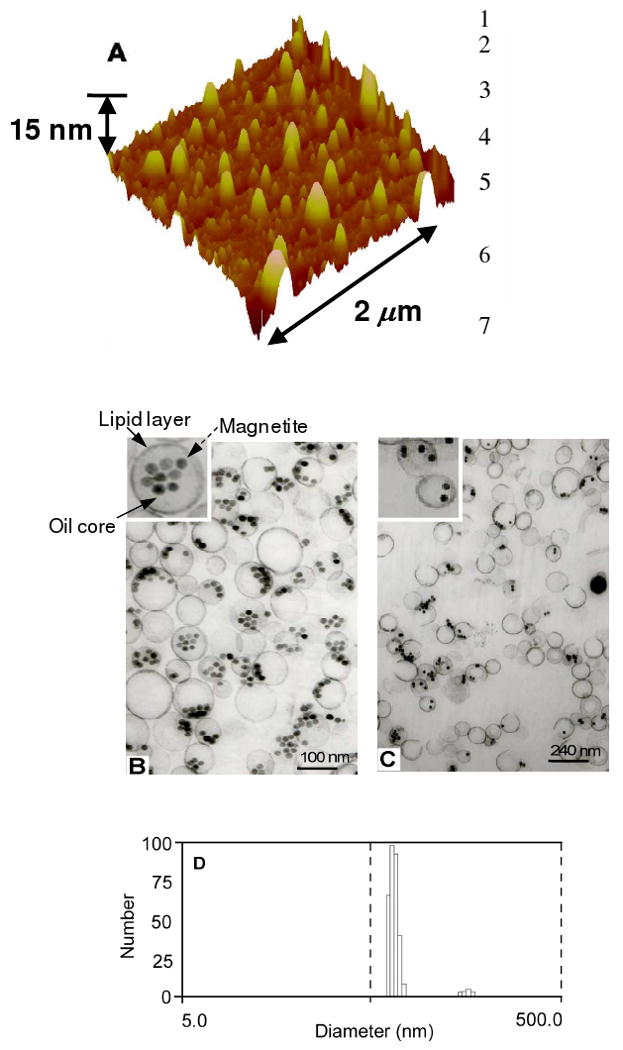
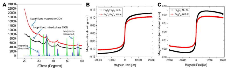
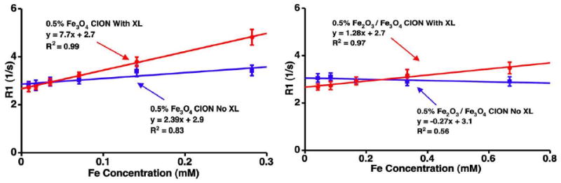
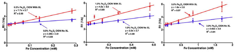
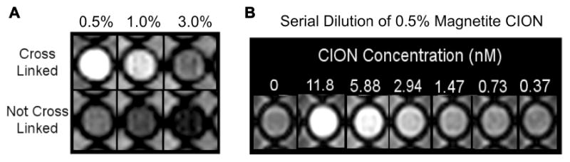
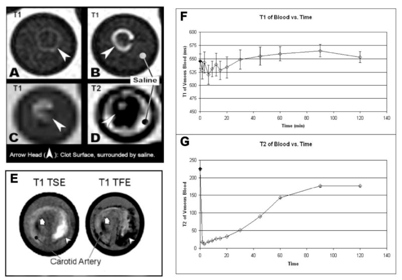
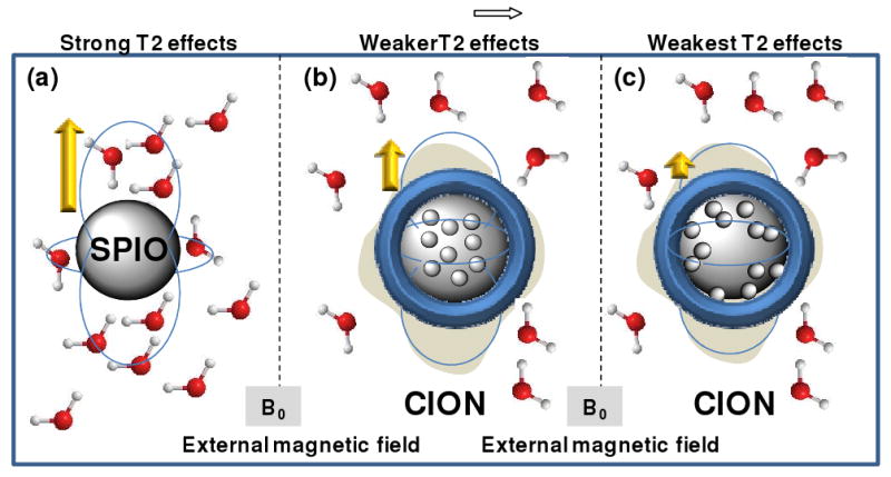
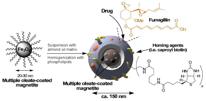
Nanomedicine approaches to atherosclerotic disease will have significant impact on the practice and outcomes of cardiovascular medicine. Iron oxide nanoparticles have been extensively used for nontargeted and targeted imaging applications based upon highly sensitive T2* imaging properties, which typically result in negative contrast effects that can only be imaged 24 or more hours after systemic administration due to persistent blood pool interference. Although recent advances involving MR pulse sequences have converted these dark contrast voxels into bright ones, the marked delays in imaging from persistent magnetic background interference and prominent dipole blooming effects of the magnetic susceptibility remain barriers to overcome. We report a T1-weighted (T1w) theranostic colloidal iron oxide nanoparticle platform, CION, which is achieved by entrapping oleate-coated magnetite particles within a cross-linked phospholipid nanoemulsion. Contrary to expectations, this formulation decreased T2 effects thus allowing positive T1w contrast detection down to low nanomolar concentrations. CION, a vascular constrained nanoplatform administered in vivo permitted T1w molecular imaging 1 h after treatment without blood pool interference, although some T2 shortening effects on blood, induced by the superparamagnetic particles, persisted. Moreover, CION was shown to encapsulate antiangiogenic drugs, like fumagillin, and retained them under prolonged dissolution, suggesting significant theranostic functionality. Overall, CION is a platform technology, developed with generally recognized as safe components, that overcomes the temporal and spatial imaging challenges associated with current iron oxide nanoparticle T2 imaging agents and which has theranostic potential in vascular diseases for detecting unstable ruptured plaque or treating atherosclerotic angiogenesis.
Figures
Similar articles
-
Controlled aggregates of magnetite nanoparticles for highly sensitive MR contrast agent.J Nanosci Nanotechnol. 2009 Dec;9(12):7118-22. doi: 10.1166/jnn.2009.1605. J Nanosci Nanotechnol. 2009. PMID: 19908740
-
Silicon nanoparticles as hyperpolarized magnetic resonance imaging agents.ACS Nano. 2009 Dec 22;3(12):4003-8. doi: 10.1021/nn900996p. ACS Nano. 2009. PMID: 19950973 Free PMC article.
-
Synthesis Of PEG-Coated, Ultrasmall, Manganese-Doped Iron Oxide Nanoparticles With High Relaxivity For T1/T2 Dual-Contrast Magnetic Resonance Imaging.Int J Nanomedicine. 2019 Oct 24;14:8499-8507. doi: 10.2147/IJN.S219749. eCollection 2019. Int J Nanomedicine. 2019. PMID: 31695377 Free PMC article.
-
Chemical synthesis and assembly of uniformly sized iron oxide nanoparticles for medical applications.Acc Chem Res. 2015 May 19;48(5):1276-85. doi: 10.1021/acs.accounts.5b00038. Epub 2015 Apr 29. Acc Chem Res. 2015. PMID: 25922976 Review.
-
Cellular magnetic resonance imaging: potential for use in assessing aspects of cardiovascular disease.Cytotherapy. 2008;10(6):575-86. doi: 10.1080/14653240802165699. Cytotherapy. 2008. PMID: 18608350 Free PMC article. Review.
Cited by
-
Nanoparticles in magnetic resonance imaging: from simple to dual contrast agents.Int J Nanomedicine. 2015 Mar 6;10:1727-41. doi: 10.2147/IJN.S76501. eCollection 2015. Int J Nanomedicine. 2015. PMID: 25834422 Free PMC article. Review.
-
Superparamagnetic iron oxide nanoparticles with variable size and an iron oxidation state as prospective imaging agents.Langmuir. 2013 Jan 15;29(2):710-6. doi: 10.1021/la3037007. Epub 2013 Jan 4. Langmuir. 2013. PMID: 23249219 Free PMC article.
-
Molecular imaging of atherosclerosis with nanoparticle-based fluorinated MRI contrast agents.Nanomedicine (Lond). 2015;10(11):1817-32. doi: 10.2217/nnm.15.26. Nanomedicine (Lond). 2015. PMID: 26080701 Free PMC article. Review.
-
Carbon-coated iron oxide nanoparticles as contrast agents in magnetic resonance imaging.Nanoscale Res Lett. 2012 Jan 5;7(1):44. doi: 10.1186/1556-276X-7-44. Nanoscale Res Lett. 2012. PMID: 22221912 Free PMC article.
-
Superparamagnetic Nanoparticles for Atherosclerosis Imaging.Nanomaterials (Basel). 2014 Jun 5;4(2):408-438. doi: 10.3390/nano4020408. Nanomaterials (Basel). 2014. PMID: 28344230 Free PMC article. Review.
References
-
- Stark DD, Weissleder R, Elizondo G, Hahn PF, Saini S, Todd LE, Wittenberg J, Ferrucci JT. Superparamagnetic Iron Oxide: Clinical Application as a Contrast Agent for MR Imaging of the Liver. Radiology. 1988;168:297–301. - PubMed
-
- Weissleder R, Hahn PF, Stark DD, Elizondo G, Saini S, Todd LE, Wittenberg J, Ferrucci JT. Superparamagnetic Iron Oxide: Enhanced Detection of Focal Splenic Tumors with MR Imaging. Radiology. 1988;169:399–403. - PubMed
-
- Frank H, Weissleder R, Brady TJ. Enhancement of MR Angiography with Iron Oxide: Preliminary Studies in Whole-Blood Phantom and In Animals. AJR Am J Roentgenol. 1994;162:209–13. - PubMed
-
- Kresse M, Wagner S, Pfefferer D, Lawaczeck R, Elste V, Semmler W. Targeting of Ultrasmall Superparamagnetic Iron Oxide (USPIO) Particles Tumor Cells In vivo by Using Transferrin Receptor Pathways. Magn Reson Med. 1998;40:236–42. - PubMed
-
- Jung C, Jacobs P. Physical and Chemical Properties of Superparamagnetic Iron Oxide MR Contrast Agents: Ferumoxides, Ferumoxtran, Ferumoxsil. Magnetic Resonance Imaging. 1995;13:661–674. - PubMed
Publication types
MeSH terms
Substances
Grants and funding
LinkOut - more resources
Full Text Sources
Other Literature Sources
Medical
