. 2009 Dec;119(12):3739-51.
doi: 10.1172/JCI39335. Epub 2009 Nov 9.

Monocyte/macrophage androgen receptor suppresses cutaneous wound healing in mice by enhancing local TNF-alpha expression

Affiliations

Monocyte/macrophage androgen receptor suppresses cutaneous wound healing in mice by enhancing local TNF-alpha expression

Jiann-Jyh Lai et al. J Clin Invest. 2009 Dec.

Abstract

Cutaneous wounds heal more slowly in elderly males than in elderly females, suggesting a role for sex hormones in the healing process. Indeed, androgen/androgen receptor (AR) signaling has been shown to inhibit cutaneous wound healing. AR is expressed in several cell types in healing skin, including keratinocytes, dermal fibroblasts, and infiltrating macrophages, but the exact role of androgen/AR signaling in these different cell types remains unclear. To address this question, we generated and studied cutaneous wound healing in cell-specific AR knockout (ARKO) mice. General and myeloid-specific ARKO mice exhibited accelerated wound healing compared with WT mice, whereas keratinocyte- and fibroblast-specific ARKO mice did not. Importantly, the rate of wound healing in the general ARKO mice was dependent on AR and not serum androgen levels. Interestingly, although dispensable for wound closure, keratinocyte AR promoted re-epithelialization, while fibroblast AR suppressed it. Further analysis indicated that AR suppressed wound healing by enhancing the inflammatory response through a localized increase in TNF-alpha expression. Furthermore, AR enhanced local TNF-alpha expression via multiple mechanisms, including increasing the inflammatory monocyte population, enhancing monocyte chemotaxis by upregulating CCR2 expression, and enhancing TNF-alpha expression in macrophages. Finally, targeting AR by topical application of a compound (ASC-J9) that degrades AR protein resulted in accelerated healing, suggesting a potential new therapeutic approach that may lead to better treatment of wound healing.


Figures

Figure 1
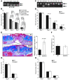
Figure 1. Cutaneous wound healing is accelerated in male GARKO mice due to loss of AR function.
Excisional wounds were made by 4-mm-diameter punches. (A) Photographs of day 8 wounds. The arrowheads point out the location of wounds. (B) Wound areas were quantified for WT and GARKO mice. n = 7–10 (2 wounds/mouse). *P = 0.001. (C) H&E staining for day 3 wound sections. Black arrowheads indicate edges of wounds, red arrowheads indicate edges of epithelium, and white arrowheads indicate foci of infiltrating cells. Scale bars: 500 μm. (D) Higher magnification of the boxed region in C. The red arrowhead indicates the edge of epithelium. Scale bar: 100 μm. (E) The quantified data for re-epithelialization. n = 4 (6 wounds/mouse). *P = 0.001. (F) Day 10 wounds were subjected to Mason’s Trichrome staining to detect collagen fibers (blue). Arrowheads indicate the edges of wounds, and ovals indicate the areas of granulation tissue. Data are representative of 12–16 wounds (4 wounds/mouse) in each group. Scale bars: 500 μm. The quantitative data show relative collagen deposition in granulation tissues compared with the adjacent dermis. n = 3–4 (4 wounds/mouse). *P < 0.05. (G) DHT pellets or placebos were implanted into GARKO mice, and only placebo pellets were implanted into WT mice. Serum DHT levels were detected by ELISA after DHT restoration. n = 3–4 mice. *P = 0.023–0.025 versus GARKO placebo. (H) After DHT restoration, wounds were created, and wound areas were quantified on the days indicated. n = 3–4 (2 wounds/mouse). *P < 0.01 versus WT (placebo).
Figure 2
Figure 2. AR in monocytes/macrophages is critical for suppressing wound healing.
(A) γ-Irradiated 6-week-old WT and GARKO mice were adoptively transplanted with WT or GARKO bone marrow cells. To confirm the bone marrow engraftment, various tissues from a WT recipient receiving GARKO bone marrow were subjected to AR genotyping. KO, truncated AR allele. (B) After bone marrow engraftment, wounds were created and the wound areas were quantified and compared among recipients. n = 3–4 (6 wounds/mouse). The numbers indicate the P values for each comparison. (C) Various tissues from MARKO mice were subjected to AR genotyping to confirm the efficiency and specificity of AR deletion. D3W, day 3 wound; Thy, thymus; Spln, spleen; Mus, muscle; Tes, testis; Bld, bladder. (D) Wound areas were compared between male MARKO mice and their WT littermates. n = 6–7 (2 wounds/mouse). (E) Day 8 wound tissues from WT and MARKO mice were subjected to Trichrome staining to detect collagen fibers (blue). Ovals indicate granulation tissues. Scale bars: 500 μm. The quantitative data show relative collagen deposition in granulation tissues compared with the adjacent dermis. n = 3 (4 wounds/mouse). (F) Day 3 wound tissues from WT and MARKO mice were subjected to H&E staining for analysis of re-epithelialization. n = 4 (6 wounds/mouse). (G) Excisional wounds were created and compared between KARKO mice and their WT littermates. n = 3–4 (6 wounds/mouse). (H) Excisional wounds were created and compared between FARKO mice and their WT littermates. n = 4–6 (6 wounds/mouse). Due to differences in genetic backgrounds among conditional ARKO lines, the WT controls among different experiments were not necessarily equivalent.
Figure 3
Figure 3. AR in keratinocytes and fibroblasts is involved in regulating re-epithelialization.
(A) Day 4 wounds from KARKO and WT mice were subjected to paraffin sectioning and H&E staining to determine the re-epithelialization rate. n = 4–6 (6 wounds/mouse). (B and C) Day 3 wounds were harvested from mice that had been injected with BrdU (150 μg/g body weight) 2 hours before harvest. The wounds were subjected to paraffin sectioning and subsequent immunohistochemistry to detect BrdU incorporation. BrdU+ epithelial cells in each wound section of (B) KARKO and (C) GARKO mice and their WT littermates were counted. n = 5–6 (6 wounds/mouse). (D) Re-epithelialization rates of day 3 wounds were compared between FARKO and WT mice. n = 4–6 (6 wounds/mouse). (E) Day 3 wounds were harvested from FARKO and WT mice injected with BrdU and subjected to immunohistochemistry as described in B and C. The proliferating (BrdU+) epithelial cells were counted in each wound section. n = 4–6 (4 wounds/mouse).
Figure 4
Figure 4. TNF-α is critical for mediating the ability of AR to suppress wound healing.
(A) Relative mRNA expression of various inflammatory mediators was measured by quantitative RT-PCR in day 3 wound tissues of WT and GARKO mice. n = 4–8. (B) Proteins were extracted from the wounded (day 3 and day 7) or non-wounded skin (control) and subjected to ELISA to detect the expression of inflammatory mediators. The concentration of TGF-β1 was measured without (active) or with (total) acid activation. n = 4–7. (C) TNF-α expression was detected by ELISA in day 3 wound tissues of MARKO mice and their WT littermates. n = 4. (D) One day after creating wounds, various concentrations of TNF-α were intradermally injected around GARKO wounds, while WT wounds were injected with PBS. The wound areas were quantified on days 4, 6, and 8. n = 3–4 (2 wounds/mouse). #P < 0.05, *P < 0.01 versus GARKO (PBS) group. (E) γ-Irradiated 6-week-old TNF-R1KO recipient mice were transplanted with bone marrow cells from WT or GARKO mice. After engraftment, wounds were created and the wound areas were quantified. n = 3–4 (6 wounds/mouse).
Figure 5
Figure 5. AR enhances local TNF-α expression through multiple mechanisms.
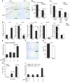
(A) Immunohistochemistry to identify infiltrating myeloid cells (CD11b+) and macrophages (F4/80+) in day 3 wounds. White arrows indicate foci of positive-staining cells (brown). Scale bars: 100 μm. (B) The positive-staining cells were counted in the wound areas. n = 6–7. (C) Flow cytometry to identify the monocytes (CD11b+F4/80+), inflammatory monocytes (CD11b+F4/80+Gr1+), and resident monocytes (CD11b+F4/80+Gr1) in PBMCs. n = 4–9. (D) mRNA isolated from BMMacs was subjected to quantitative RT-PCR to detect expression of chemokine receptors. For each chemokine receptor, the expression level of WT BMMacs treated with LPS was set as 1. (E) NIH3T3 cells were transiently transfected with reporter containing the promoter region (–1,258 to +27) of murine CCR2 together with pWPI-mAR or empty vector. After treating cells with or without DHT, luciferase activities were measured. (F and G) Transwell chemotaxis assay was used to compare chemotaxis of day 3 BMMacs in response to MCP-1. (F) Images of cells that had migrated across the membrane. Original magnification, ×100. (G) Cell numbers were counted. n = 9. (H) Day 6 BMMacs were treated with LPS as indicated. TNF-α production was measured by ELISA. (I) Reporters composed of TNF-α promoter containing 5′-UTR (pGL3-TNF, –1,203 to +122) or without 5′-UTR (pGL3-TNF, –1,203 to +2) were cotransfected into NIH3T3 cells with pWPI-mAR or pWPI. After treating with DHT, luciferase activities were measured. Data in I are mean ± SD. *P < 0.001 versus all other groups.
Figure 6
Figure 6. ASC-J9 accelerates cutaneous wound healing in WT mice and suppresses TNF-α production of wounds and BMMacs.
(A) ASC-J9 cream (50 μM) was applied topically on the wounds of WT mice daily, from day 0 to day 8. The wound areas were quantified to compare the effects of ASC-J9 versus the vehicle cream. n = 3–4 (6 wounds/mouse). (B) TNF-α protein expression was detected by ELISA in day 3 and day 7 wound tissues from ASC-J9– or vehicle-treated mice (pool 6 wounds/mouse in 0.7 ml homogenization buffer). n = 4. (C) Day 6 BMMacs (8 × 10⁴/well) were cultured in RPMI-CD medium overnight, then treated with 1 μM ASC-J9 or DMSO for the entire culture period. Four hours after starting ASC-J9 treatment, 1 nM DHT or ethanol was added into the culture for another 4 hours, followed by 1 ng/ml LPS plus 1 μM ASC-J9 or DMSO for 24 hours. TNF-α production in the supernatant was measured by ELISA.
Figure 7
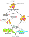
Figure 7. AR enhances local TNF-α expression from macrophages through multiple mechanisms to suppress cutaneous wound healing.
Monocyte/macrophage AR function can be modulated by androgens or other factors to enhance inflammatory responses induced by injury. Overall, local TNF-α expression from macrophages is augmented by AR and critically mediates wound-healing suppression by targeting resident skin cells (keratinocytes and/or fibroblasts) via TNF-R1. AR enhances local TNF-α expression through 3 mechanisms. First, AR increases the circulating inflammatory monocyte population. Second, AR promotes the expression of CCR2 on inflammatory monocytes to enhance their chemotaxis in response to MCP-1. Third, AR can directly enhance TNF-α expression of macrophages in response to LPS stimulation. Treatment with ASC-J9 can antagonize the function of AR and promote cutaneous wound healing.

Similar articles

Cited by

References

    1. Ashcroft G.S., Mills S.J. Androgen receptor-mediated inhibition of cutaneous wound healing. J. Clin. Invest. 2002;110:615–624. - PMC - PubMed
    1. Ashcroft G.S., Greenwell-Wild T., Horan M.A., Wahl S.M., Ferguson M.W. Topical estrogen accelerates cutaneous wound healing in aged humans associated with an altered inflammatory response. Am. J. Pathol. 1999;155:1137–1146. - PMC - PubMed
    1. Fimmel S., Zouboulis C.C. Influence of physiological androgen levels on wound healing and immune status in men. Aging Male. 2005;8:166–174. doi: 10.1080/13685530500233847. - DOI - PubMed
    1. Taylor R.J., Taylor A.D., Smyth J.V. Using an artificial neural network to predict healing times and risk factors for venous leg ulcers. J. Wound Care. 2002;11:101–105. - PubMed
    1. Chen W., et al. Evidence of heterogeneity and quantitative differences of the type 1 5alpha-reductase expression in cultured human skin cells — evidence of its presence in melanocytes. J. Invest. Dermatol. 1998;110:84–89. doi: 10.1046/j.1523-1747.1998.00080.x. - DOI - PubMed

Publication types

MeSH terms

Substances