

Synovial fibroblasts spread rheumatoid arthritis to unaffected joints
- PMID: 19898488
- PMCID: PMC3678354
- DOI: 10.1038/nm.2050
Synovial fibroblasts spread rheumatoid arthritis to unaffected joints
Abstract
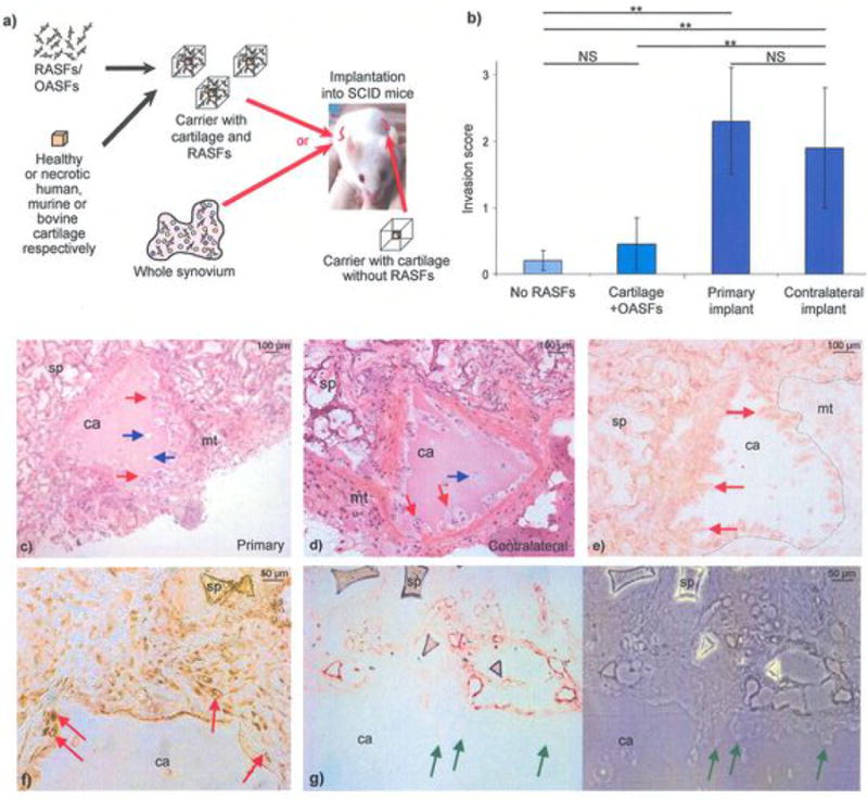
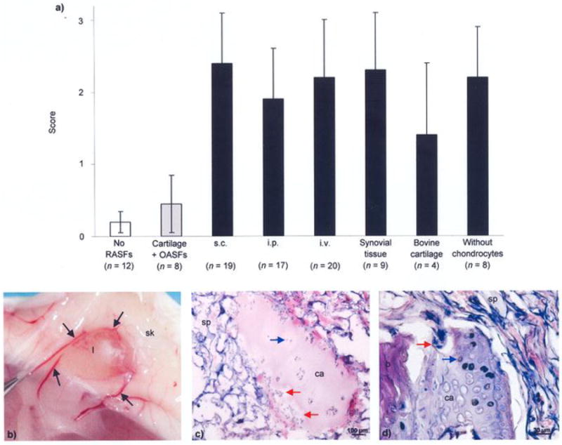
Active rheumatoid arthritis originates from few joints but subsequently affects the majority of joints. Thus far, the pathways of the progression of the disease are largely unknown. As rheumatoid arthritis synovial fibroblasts (RASFs) which can be found in RA synovium are key players in joint destruction and are able to migrate in vitro, we evaluated the potential of RASFs to spread the disease in vivo. To simulate the primary joint of origin, we implanted healthy human cartilage together with RASFs subcutaneously into severe combined immunodeficient (SCID) mice. At the contralateral flank, we implanted healthy cartilage without cells. RASFs showed an active movement to the naive cartilage via the vasculature independent of the site of application of RASFs into the SCID mouse, leading to a marked destruction of the target cartilage. These findings support the hypothesis that the characteristic clinical phenomenon of destructive arthritis spreading between joints is mediated, at least in part, by the transmigration of activated RASFs.
Conflict of interest statement
Competing interests: The authors have no competing interests.
Figures

Similar articles
-
Migratory potential of rheumatoid arthritis synovial fibroblasts: additional perspectives.Cell Cycle. 2010 Jun 15;9(12):2286-91. doi: 10.4161/cc.9.12.11907. Epub 2010 Jun 15. Cell Cycle. 2010. PMID: 20519953
-
Cartilage destruction mediated by synovial fibroblasts does not depend on proliferation in rheumatoid arthritis.Am J Pathol. 2003 May;162(5):1549-57. doi: 10.1016/S0002-9440(10)64289-7. Am J Pathol. 2003. PMID: 12707039 Free PMC article.
-
Retroviral gene transfer of an antisense construct against membrane type 1 matrix metalloproteinase reduces the invasiveness of rheumatoid arthritis synovial fibroblasts.Arthritis Rheum. 2005 Jul;52(7):2010-4. doi: 10.1002/art.21156. Arthritis Rheum. 2005. PMID: 15986375
-
Rheumatoid arthritis progression mediated by activated synovial fibroblasts.Trends Mol Med. 2010 Oct;16(10):458-68. doi: 10.1016/j.molmed.2010.07.004. Epub 2010 Aug 24. Trends Mol Med. 2010. PMID: 20739221 Review.
-
Synovial fibroblasts: key players in rheumatoid arthritis.Rheumatology (Oxford). 2006 Jun;45(6):669-75. doi: 10.1093/rheumatology/kel065. Epub 2006 Mar 27. Rheumatology (Oxford). 2006. PMID: 16567358 Review.
Cited by
-
Celastrol inhibits lipopolysaccharide-stimulated rheumatoid fibroblast-like synoviocyte invasion through suppression of TLR4/NF-κB-mediated matrix metalloproteinase-9 expression.PLoS One. 2013 Jul 4;8(7):e68905. doi: 10.1371/journal.pone.0068905. Print 2013. PLoS One. 2013. PMID: 23861949 Free PMC article.
-
Toll-like receptor 2 (TLR2) induces migration and invasive mechanisms in rheumatoid arthritis.Arthritis Res Ther. 2015 Jun 9;17(1):153. doi: 10.1186/s13075-015-0664-8. Arthritis Res Ther. 2015. PMID: 26055925 Free PMC article.
-
Adipokines and Inflammation Alter the Interaction Between Rheumatoid Arthritis Synovial Fibroblasts and Endothelial Cells.Front Immunol. 2020 Jun 2;11:925. doi: 10.3389/fimmu.2020.00925. eCollection 2020. Front Immunol. 2020. PMID: 32582145 Free PMC article.
-
IL-26 is overexpressed in rheumatoid arthritis and induces proinflammatory cytokine production and Th17 cell generation.PLoS Biol. 2012;10(9):e1001395. doi: 10.1371/journal.pbio.1001395. Epub 2012 Sep 25. PLoS Biol. 2012. PMID: 23055831 Free PMC article.
-
A protocol for humanized synovitis mice model.Am J Clin Exp Immunol. 2019 Oct 15;8(5):47-52. eCollection 2019. Am J Clin Exp Immunol. 2019. PMID: 31777685 Free PMC article.
References
-
- Karouzakis E, Neidhart M, Gay RE, Gay S. Molecular and cellular basis of rheumatoid joint destruction. Immunol Lett. 2006;106:8–13. - PubMed
-
- Muller-Ladner U, Pap T, Gay RE, Neidhart M, Gay S. Mechanisms of disease: the molecular and cellular basis of joint destruction in rheumatoid arthritis. Nat Clin Pract Rheumatol. 2005;1:102–10. - PubMed
-
- Looney RJ. B cell-targeted therapy for rheumatoid arthritis: an update on the evidence. Drugs. 2006;66:625–39. - PubMed
Publication types
MeSH terms
Grants and funding
LinkOut - more resources
Full Text Sources
Other Literature Sources
Medical
Molecular Biology Databases
Research Materials
