. 2010 Jan 8;285(2):1516-28.
doi: 10.1074/jbc.M109.028241. Epub 2009 Nov 5.

The vacuole import and degradation pathway utilizes early steps of endocytosis and actin polymerization to deliver cargo proteins to the vacuole for degradation

Affiliations

The vacuole import and degradation pathway utilizes early steps of endocytosis and actin polymerization to deliver cargo proteins to the vacuole for degradation

C Randell Brown et al. J Biol Chem. .

Abstract

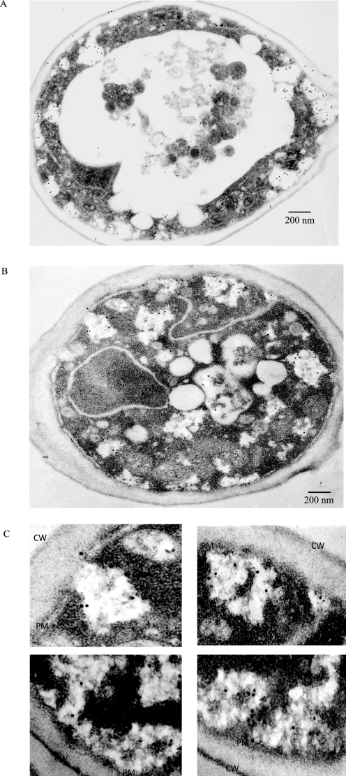
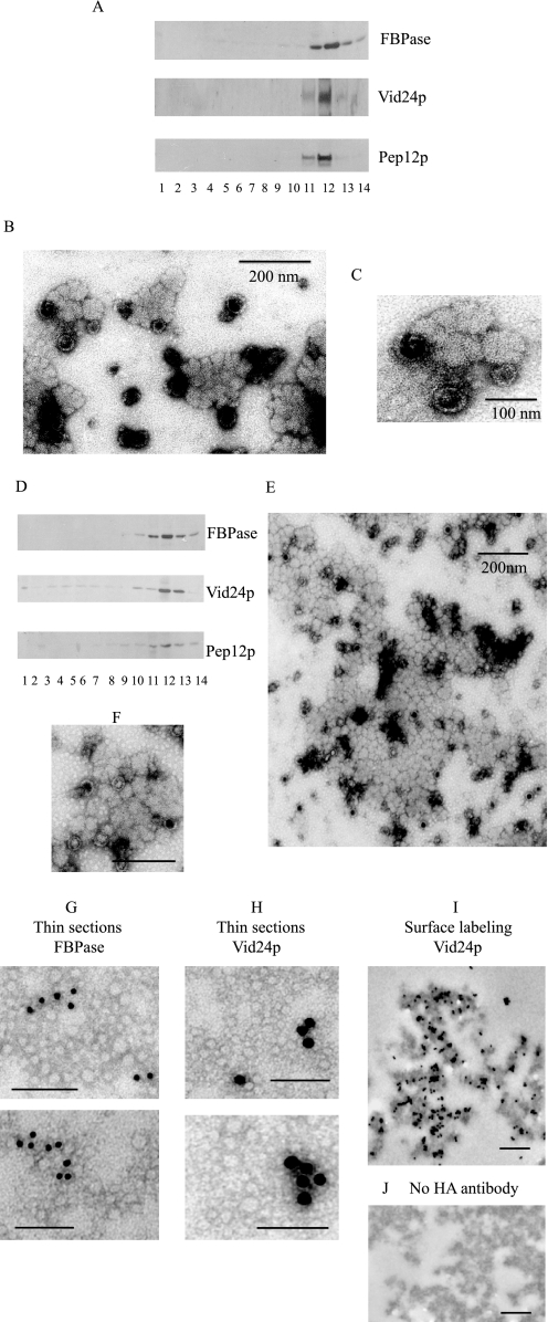
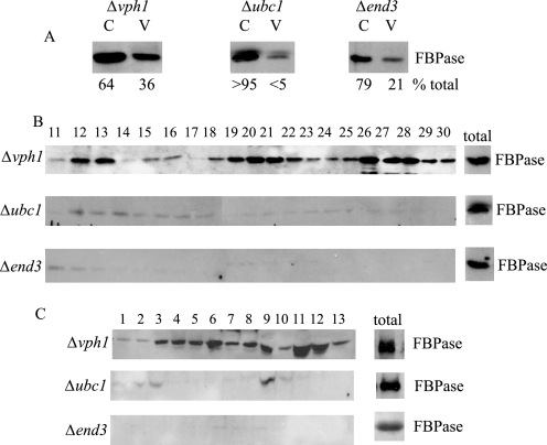
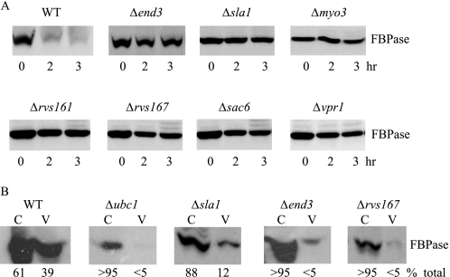
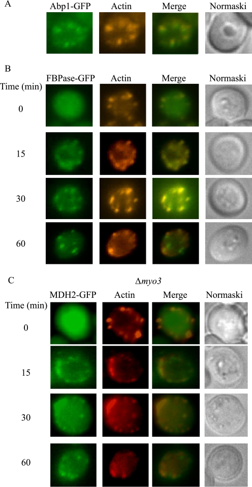
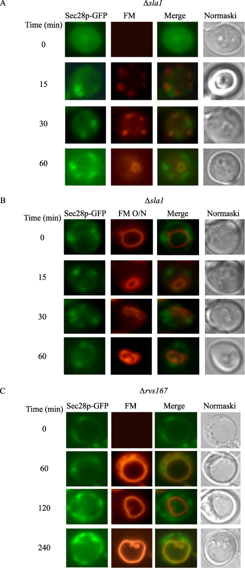
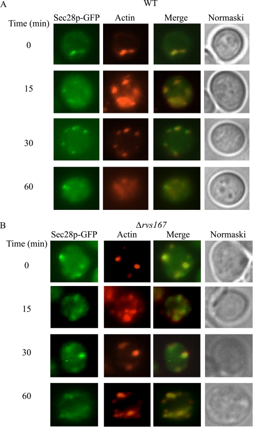
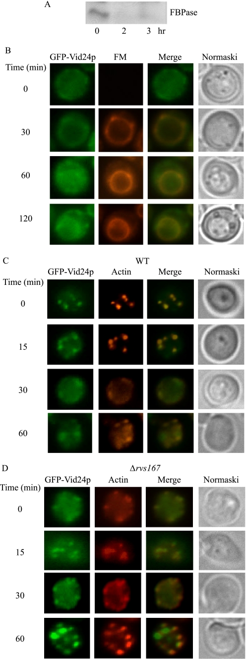
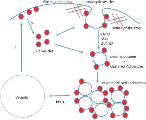
When glucose is added to yeast cells that are starved for 3 days, fructose-1,6-bisphosphatase (FBPase) and malate dehydrogenase 2 are degraded in the vacuole via the vacuole import and degradation (Vid) pathway. In this study, we examined the distribution of FBPase at the ultrastructural level. FBPase was observed in areas close to the plasma membrane and in cytoplasmic structures that are heterogeneous in size and density. We have isolated these intracellular structures that contain FBPase, the Vid vesicle marker Vid24p, and the endosomal marker Pep12p. They appeared irregular in size and shape. In yeast, actin polymerization plays an important role in early steps of endocytosis. Mutants that affect actin polymerization inhibited FBPase degradation, suggesting that actin polymerization is important for FBPase degradation. Both FBPase and malate dehydrogenase 2 were associated with actin patches. Vid vesicle proteins such as Vid24p or Sec28p were also at actin patches, although they dissociated from these structures at later time points. We propose that Vid24p and Sec28p are present at actin patches during glucose starvation. Cargo proteins arrive at these sites following the addition of glucose, and the endocytic vesicles then pinch off from the plasma membrane. Following the fusion of endosomes with the vacuole, cargo proteins are then degraded in the vacuole.


Figures

FIGURE 1.
FIGURE 1.
Immunoelectron microscopy studies of FBPase distribution in wild-type and Δpep4 cells following the addition of glucose. A, wild-type cells were glucose-starved and transferred to media containing fresh glucose for 15 min. Cells were fixed and embedded. Immuno-EM was performed on thin sections of cells using affinity-purified FBPase antibodies followed by goat anti-rabbit antibodies conjugated with 10 nm colloid gold. B, Δpep4 cells were glucose-starved and transferred to media containing glucose for 15 min. Cells were fixed, and immuno-EM was performed on thin sections of cells. C, enlarged views of FBPase distribution in areas near the plasma membrane. CW, cell wall; PM, plasma membrane.
FIGURE 2.
FIGURE 2.
Isolation of intracellular structures that contain FBPase, Vid24p, and Pep12p. A, wild-type cells expressing HA-Vid24p were transferred to media containing glucose for 15 min. Cells were spheroplasted and subjected to differential centrifugation. The 100,000 × g pellets were re-suspended and further fractionated over an S-1000 column. Fractions 19–22 were pooled and centrifuged at 100,000 × g. Pellets were re-suspended and subjected to two sequential 20–70% sucrose density gradients. On the second gradient, the distribution of FBPase, HA-Vid24p, and Pep12p was determined by Western blotting with anti-FBPase, anti-HA, and anti-Pep12p antibodies. B, fraction 12 from the second sucrose gradient was collected and examined by negative staining and visualized by electron microscopy. C, enlarged views of organelles isolated from fraction 12. D, fractions 15–18 were collected from the S-1000 column and subjected to two sequential sucrose density gradients. The distribution of FBPase, HA-Vid24p, and Pep12p was examined by Western blotting. E, fraction 12 was collected and examined by electron microscopy using negative staining. F, enlarged views of FBPase-containing structures purified from fraction 12. Bars equal 200 nm. G, FBPase-containing organelles were purified, fixed, and embedded in LR White. Thin sections were incubated with anti-FBPase antibodies followed by goat anti-rabbit secondary antibodies conjugated with 10 nm gold particles. H, thin sections were incubated with anti-HA antibodies followed by goat anti-mouse secondary antibodies conjugated with 15 nm gold particles. I, FBPase-containing organelles were purified, fixed, and incubated with anti-HA antibodies followed by anti-mouse antibodies conjugated with 10 nm gold particles. J, control samples were processed the same way as in I but in the absence of anti-HA antibodies. Bars equal 200 nm.
FIGURE 3.
FIGURE 3.
FBPase levels in the Vid vesicle fraction are reduced in Δend3 mutants that block early steps of endocytosis. A, cells lacking VPH1, UBC1, and END3 were shifted to glucose for 15 min, and cell lysates were subjected to differential centrifugation. FBPase distribution in the Vid vesicle-enriched fraction (V, 200,000 × g pellet) and cytosolic fraction (C, 200,000 × g supernatant) was examined by Western blotting with anti-FBPase antibodies. Levels of FBPase in C and V fractions were quantitated by NIH ImageJ software. B, the same cells were transferred to media containing fresh glucose for 15 min and subjected to differential centrifugation. The 100,000 × g pellets were re-suspended and further separated by S-1000 columns. Fractions were collected, and the distribution of FBPase was examined. C, the same cells were glucose-starved and re-fed with fresh glucose for 15 min. Cell lysates were centrifuged by differential centrifugation. The 100,000 × g pellets were re-suspended and loaded onto 20–70% sucrose density gradients. Fractions were precipitated by trichloroacetic acid, and the distribution of FBPase was examined.
FIGURE 4.
FIGURE 4.
FBPase degradation is defective in mutants that block early steps of endocytosis. A, wild-type and various mutant cells that block early steps of endocytosis were grown in low glucose media. Cells were transferred to media containing fresh glucose for 0, 2, and 3 h, and FBPase degradation was examined. B, wild-type and endocytosis mutants were transferred from low to high glucose for 15 min. Cell lysates were subjected to differential centrifugation and FBPase distribution in the cytosolic (C) and Vid vesicle (V)-enriched fractions was examined. FBPase levels in these fractions were quantitated using ImageJ software (National Institutes of Health).
FIGURE 5.
FIGURE 5.
FBPase remains in the cytosol when early steps of endocytosis are blocked. The Δvph1 cell expressing FBPase-GFP (A), Δsla1 cells expressing FBPase-GFP (B), and Δrvs167 cells expressing FBPase-GFP (C) were grown in low glucose media. Cells were transferred to media containing fresh glucose in the presence of FM for the indicated times. FBPase-GFP, FM, and cells were visualized using fluorescence microscopy.
FIGURE 6.
FIGURE 6.
FBPase and MDH2 associate with actin patches following the addition of glucose. A, wild-type cells expressing Abp1p-GFP were grown in YPKG media for 3 days. The localization of Abp1p-GFP with rhodamine-conjugated phalloidin was examined. B, wild-type cells expressing FBPase-GFP were transferred to media containing fresh glucose for the indicated times. Cells were fixed and processed as described under “Experimental Procedures.” Actin was stained with phalloidin conjugated with rhodamine. FBPase, actin, and cells were visualized by fluorescence microscopy. C, MDH2-GFP was expressed in Δmyo3 cells that were transferred to media containing glucose for 0–60 min. MDH2 and actin localization was examined by fluorescence microscopy.
FIGURE 7.
FIGURE 7.
Sec28p is present at the cell periphery in the Δsla1 and Δrvs167mutants. A, Sec28p-GFP was expressed in Δsla1 cells. These cells were re-fed with glucose in the presence of FM. Sec28p localization with FM was examined. B, Δsla1 cells were incubated with FM for 16 h. Cells were then shifted to glucose in the absence of FM. The distribution of Sec28p and FM was examined. C, cells lacking the RVS167 gene were transformed with Sec28p-GFP. Cells were re-fed with glucose in the presence of FM for the indicated times. The distribution of Sec28p and FM was examined.
FIGURE 8.
FIGURE 8.
Sec28p co-localizes with actin patches in wild-type cells. A, wild-type cells expressing Sec28p-GFP were transferred to media containing fresh glucose for 0–60 min. The distribution of Sec28p and actin patches was examined. B, the Δrvs167 mutant was transformed with Sec28p-GFP and grown in low glucose media. Cells were transferred to media containing fresh glucose for the indicated time. The distribution of Sec28p and actin patches was examined.
FIGURE 9.
FIGURE 9.
Vid24p co-localizes with actin patches in wild-type cells. A, GFP-Vid24p was transformed into wild-type cells that were grown in low glucose media for 3 days and re-fed with glucose for 0, 2, and 3 h. FBPase degradation was examined. B, wild-type cells expressing GFP-Vid24p were transferred to media containing fresh glucose in the presence of FM for the indicated times. Vid24p distribution with FM was examined. C, wild-type cells expressing GFP-Vid24p were glucose-starved and then transferred to glucose containing media. The distribution of GFP-Vid24p with actin patches was examined by fluorescence microscopy. D, GFP-Vid24p was expressed in Δrvs167 cells that were grown in low glucose media. Glucose was then added to these cells for 0–60 min. Vid24p and actin patches were examined using fluorescence microscopy.
FIGURE 10.
FIGURE 10.
The Vid pathway utilizes a specialized endocytic pathway. During glucose starvation, Vid vesicle proteins are present at actin patches where endocytic vesicles are forming from the plasma membrane. Following the addition of glucose, cargo proteins arrive at these actin patches. Endocytic vesicles use actin polymerization to pinch off from the plasma membrane. Small endosomes that are released from the plasma membrane may cluster or fuse together to form large endosomes. These large endosomes then fuse with the vacuole. Vid vesicles may attach to endocytic vesicles that are forming from the plasma membrane. Vid vesicles may continue to attach to endosomes that are released into the cytoplasm. It is possible that Vid vesicles fuse directly with the vacuole. We suggest that Vid vesicles are formed from different regions of the plasma membrane. They may be derived from retrograde vesicles coming from the vacuole membrane. These retrograde vesicles travel to the plasma membrane and then become Vid vesicles.

Similar articles

Cited by

References

    1. Goldberg A. L. (2007) Biochem. Soc. Trans. 35, 12–17 - PubMed
    1. Hanna J., Finley D. (2007) FEBS Lett. 581, 2854–2861 - PMC - PubMed
    1. Varshavsky A. (2005) Trends Biochem. Sci. 30, 283–286 - PubMed
    1. Seglen P. O., Gordon P. B., Holen I. (1990) Semin. Cell Biol. 1, 441–448 - PubMed
    1. Neely A. N., Cox J. R., Fortney J. A., Schworer C. M., Mortimore G. E. (1977) J. Biol. Chem. 252, 6948–6954 - PubMed

Publication types

MeSH terms

LinkOut - more resources