Inflammatory bowel disease and mutations affecting the interleukin-10 receptor
- PMID: 19890111
- PMCID: PMC2787406
- DOI: 10.1056/NEJMoa0907206
Inflammatory bowel disease and mutations affecting the interleukin-10 receptor
Abstract
Background: The molecular cause of inflammatory bowel disease is largely unknown.
Methods: We performed genetic-linkage analysis and candidate-gene sequencing on samples from two unrelated consanguineous families with children who were affected by early-onset inflammatory bowel disease. We screened six additional patients with early-onset colitis for mutations in two candidate genes and carried out functional assays in patients' peripheral-blood mononuclear cells. We performed an allogeneic hematopoietic stem-cell transplantation in one patient.
Results: In four of nine patients with early-onset colitis, we identified three distinct homozygous mutations in genes IL10RA and IL10RB, encoding the IL10R1 and IL10R2 proteins, respectively, which form a heterotetramer to make up the interleukin-10 receptor. The mutations abrogate interleukin-10-induced signaling, as shown by deficient STAT3 (signal transducer and activator of transcription 3) phosphorylation on stimulation with interleukin-10. Consistent with this observation was the increased secretion of tumor necrosis factor alpha and other proinflammatory cytokines from peripheral-blood mononuclear cells from patients who were deficient in IL10R subunit proteins, suggesting that interleukin-10-dependent "negative feedback" regulation is disrupted in these cells. The allogeneic stem-cell transplantation performed in one patient was successful.
Conclusions: Mutations in genes encoding the IL10R subunit proteins were found in patients with early-onset enterocolitis, involving hyperinflammatory immune responses in the intestine. Allogeneic stem-cell transplantation resulted in disease remission in one patient.
2009 Massachusetts Medical Society
Figures
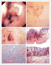
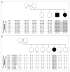
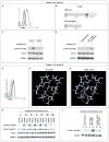
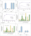
Comment in
-
Interleukin-10 in inflammatory bowel disease.N Engl J Med. 2009 Nov 19;361(21):2091-3. doi: 10.1056/NEJMe0909225. Epub 2009 Nov 4. N Engl J Med. 2009. PMID: 19890110 No abstract available.
-
Defective interleukin-10 signaling in human inflammatory bowel disease.Gastroenterology. 2010 May;138(5):2016-8. doi: 10.1053/j.gastro.2010.03.018. Epub 2010 Mar 19. Gastroenterology. 2010. PMID: 20303964 No abstract available.
-
IL-10RA truncation mutations and Semite populations.Inflamm Bowel Dis. 2011 Jun;17(6):1438. doi: 10.1002/ibd.21495. Epub 2010 Sep 14. Inflamm Bowel Dis. 2011. PMID: 20842643 No abstract available.
Similar articles
-
Loss of interleukin-10 signaling and infantile inflammatory bowel disease: implications for diagnosis and therapy.Gastroenterology. 2012 Aug;143(2):347-55. doi: 10.1053/j.gastro.2012.04.045. Epub 2012 Apr 28. Gastroenterology. 2012. PMID: 22549091
-
Clinical features of interleukin 10 receptor gene mutations in children with very early-onset inflammatory bowel disease.J Pediatr Gastroenterol Nutr. 2015 Mar;60(3):332-8. doi: 10.1097/MPG.0000000000000621. J Pediatr Gastroenterol Nutr. 2015. PMID: 25373860
-
Interleukin 1β Mediates Intestinal Inflammation in Mice and Patients With Interleukin 10 Receptor Deficiency.Gastroenterology. 2016 Dec;151(6):1100-1104. doi: 10.1053/j.gastro.2016.08.055. Epub 2016 Sep 28. Gastroenterology. 2016. PMID: 27693323 Free PMC article.
-
Candidiasis associated with very early onset inflammatory bowel disease: First IL10RB deficient case from the National Iranian Registry and review of the literature.Clin Immunol. 2019 Aug;205:35-42. doi: 10.1016/j.clim.2019.05.007. Epub 2019 May 13. Clin Immunol. 2019. PMID: 31096038 Review.
-
Interleukin-10 and interleukin-10-receptor defects in inflammatory bowel disease.Curr Allergy Asthma Rep. 2012 Oct;12(5):373-9. doi: 10.1007/s11882-012-0286-z. Curr Allergy Asthma Rep. 2012. PMID: 22890722 Review.
Cited by
-
Host-pathobiont interactions in Crohn's disease.Nat Rev Gastroenterol Hepatol. 2024 Oct 24. doi: 10.1038/s41575-024-00997-y. Online ahead of print. Nat Rev Gastroenterol Hepatol. 2024. PMID: 39448837 Review.
-
Type 17 immunity: novel insights into intestinal homeostasis and autoimmune pathogenesis driven by gut-primed T cells.Cell Mol Immunol. 2024 Nov;21(11):1183-1200. doi: 10.1038/s41423-024-01218-x. Epub 2024 Oct 8. Cell Mol Immunol. 2024. PMID: 39379604 Review.
-
Effects of breast-fed infants-derived Limosilactobacillus reuteri and Bifidobacterium breve ameliorate DSS-induced colitis in mice.iScience. 2024 Sep 11;27(10):110902. doi: 10.1016/j.isci.2024.110902. eCollection 2024 Oct 18. iScience. 2024. PMID: 39351200 Free PMC article.
-
[A case report of Crohn's disease in a child with trisomy 8].Zhongguo Dang Dai Er Ke Za Zhi. 2024 Sept 15;26(9):982-985. doi: 10.7499/j.issn.1008-8830.2405069. Zhongguo Dang Dai Er Ke Za Zhi. 2024. PMID: 39267515 Free PMC article. Chinese.
-
Successful Allogeneic Hematopoietic Cell Transplantation for Patients with IL10RA Deficiency in Japan.J Clin Immunol. 2024 Sep 12;45(1):6. doi: 10.1007/s10875-024-01795-6. J Clin Immunol. 2024. PMID: 39264505
References
-
- Podolsky DK. Inflammatory bowel disease. N Engl J Med. 2002;347:417–29. - PubMed
-
- Xavier RJ, Podolsky DK. Unravelling the pathogenesis of inflammatory bowel disease. Nature. 2007;448:427–34. - PubMed
-
- Fried K, Vure E. A lethal autosomal recessive enterocolitis of early infancy. Clin Genet. 1974;6:195–6. - PubMed
-
- Mégarbané A, Sayad R. Early lethal autosomal recessive enterocolitis: report of a second family. Clin Genet. 2007;71:89–90. - PubMed
Publication types
MeSH terms
Substances
Grants and funding
LinkOut - more resources
Full Text Sources
Other Literature Sources
Molecular Biology Databases
Miscellaneous
