. 2009 Dec 1;183(11):6989-97.
doi: 10.4049/jimmunol.0901386. Epub 2009 Nov 4.

Role of double-stranded RNA pattern recognition receptors in rhinovirus-induced airway epithelial cell responses

Affiliations

Role of double-stranded RNA pattern recognition receptors in rhinovirus-induced airway epithelial cell responses

Qiong Wang et al. J Immunol. .

Abstract

Rhinovirus (RV), a ssRNA virus of the picornavirus family, is a major cause of the common cold as well as asthma and chronic obstructive pulmonary disease exacerbations. Viral dsRNA produced during replication may be recognized by the host pattern recognition receptors TLR-3, retinoic acid-inducible gene (RIG)-I, and melanoma differentiation-associated gene (MDA)-5. No study has yet identified the receptor required for sensing RV dsRNA. To examine this, BEAS-2B human bronchial epithelial cells were infected with intact RV-1B or replication-deficient UV-irradiated virus, and IFN and IFN-stimulated gene expression was determined by quantitative PCR. The separate requirements of RIG-I, MDA5, and IFN response factor (IRF)-3 were determined using their respective small interfering RNAs (siRNA). The requirement of TLR3 was determined using siRNA against the TLR3 adaptor molecule Toll/IL-1R homologous region-domain-containing adapter-inducing IFN-beta (TRIF). Intact RV-1B, but not UV-irradiated RV, induced IRF3 phosphorylation and dimerization, as well as mRNA expression of IFN-beta, IFN-lambda1, IFN-lambda2/3, IRF7, RIG-I, MDA5, 10-kDa IFN-gamma-inducible protein/CXCL10, IL-8/CXCL8, and GM-CSF. siRNA against IRF3, MDA5, and TRIF, but not RIG-I, decreased RV-1B-induced expression of IFN-beta, IFN-lambda1, IFN-lambda2/3, IRF7, RIG-I, MDA5, and inflammatory protein-10/CXCL10 but had no effect on IL-8/CXCL8 and GM-CSF. siRNAs against MDA5 and TRIF also reduced IRF3 dimerization. Finally, in primary cells, transfection with MDA5 siRNA significantly reduced IFN expression, as it did in BEAS-2B cells. These results suggest that TLR3 and MDA5, but not RIG-I, are required for maximal sensing of RV dsRNA and that TLR3 and MDA5 signal through a common downstream signaling intermediate, IRF3.


Figures
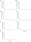
Figure 1
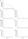
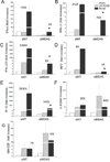
Figure 1. Fold increase of RV1B-induced IFN and ISG responses in cultured airway epithelial cells
BEAS-2B cells were infected with RV1B or sham (1 h at 33°C). Total RNA was extracted at 1, 8, 16, 24, 48 and 72 hours after infection. A–G. The expression of IFN-β, IFN-λ1, IFN-λ2/3, IRF7, RIG-I, MDA5 and TLR3 at each time point was determined by qPCR. The expression of each target gene was normalized to GAPDH. Expression levels are represented as the ratio of the response to intact RV vs. the response to sham. Data represent mean ± SEM for three experiments.
Figure 2
Figure 2
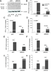
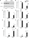
RV1B-induced protein expression of IFNs and ISGs. BEAS-2B cells were grown to near confluence and infected with RV1B, UV-irradiated RV1B or sham. A–C. Medium supernatants were extracted twenty-four hours after infection for ELISA to determine the expression of IFN-λ1, IP-10 and IL-8. D. Protein lysates were used to determine IRF7 expression by immunoblotting.
Figure 3
Figure 3. RV1B-induced expression of IFNs, ISGs and chemokines in primary tracheobronchial epithelial cells
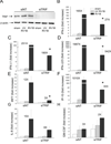
Primary tracheobronchial epithelial cells were grown to near confluence and infected with RV1B or sham. A. Total RNA was extracted twenty-four hour after infection, and the expression of IFN-β, IFN-λ1, IFN-λ2/3, IRF7, IP-10, IL-8 and GM-CSF determined by qPCR. Expression levels are represented as the ratio of the response to intact RV vs. the response to sham. The y-axis is in log scale. B–G. Time course of RV1B-induced responses in primary cells.
Figure 4
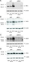
Figure 4. RIG-I and MDA-5 siRNA knockdown efficiencies
A, B. RIG-I or non-targeting siRNA was transfected into BEAS-2B cells. After transfection, cells were infected with RV1B, UV-irradiated RV1B (UV-RV1B) or sham. After infection, cell lysates were probed with antibodies against RIG-I (A) or MDA-5 (B). Note the inductions in RIG-I and MDA5 expression with intact RV, as well as the apparent degradation of MDA-5 following viral infection. C, D. MDA5 or non-targeting siRNA was transfected into BEAS-2B cells. After transfection, cells were infected with RV1B, UV-irradiated RV1B (UV-RV1B) or sham. After infection, cell lysates were probed with antibodies against either MDA5 (C) or RIG-I (D). (The blots shown are a representative of three separate experiments.)
Figure 5
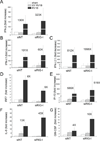
Figure 5. siRNA against RIG-I does not block RV1B-induced IFN and ISG expression
A–G. Total RNA was extracted and the expression of IFN-β, IFN-λ1, IFN-λ2/3, IRF7, IP-10, IL-8 and GM-CSF was determined by qPCR. The expression of each target gene was normalized to GAPDH. Expression levels are represented as the fold increase vs. sham-infected, non-targeting siRNA-transfected cells. The y-axis has been broken in order to show the effects of siRNA on both basal and maximal gene expression. Bars represent mean ± SEM for four experiments; numbers on top of bars indicate the fold increase compared to sham-infected sample within its own siRNA group. (*p<0.05 vs. RIG-I siRNA-transfected RV1B-infected sample, ANOVA.)
Figure 6
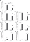
Figure 6. siRNA against MDA5 blocks RV1B-induced IFN and ISG expression
A–G. Total RNA was extracted and the expression of IFN-β, IFN-λ1, IFN-λ2/3, IRF7, IP-10, IL-8 and GM-CSF was determined by qPCR. The expression of each target gene was normalized to GAPDH. Expression levels are represented as the fold increase vs. sham-infected, non-targeting siRNA-transfected cells. The y-axis has been broken in order to show the effects of siRNA on both basal and maximal gene expression. Bars represent mean ± SEM for five experiments; numbers on top of bars indicate the fold increase compared to sham-infected sample within its own siRNA group. (*p<0.05 vs. MDA5 siRNA-transfected RV-infected sample, ANOVA.)
Figure 7
Figure 7. siRNA against MDA5 blocks RV39-induced IFN and ISG expression
A–G. Total RNA was extracted and the expression of IFN-β, IFN-λ1, IFN-λ2/3, IRF7, IP-10, IL-8 and GM-CSF was determined by qPCR. The expression of each target gene was normalized to GAPDH. Expression levels are represented as the fold increase vs. sham-infected, non-targeting siRNA-transfected cells. The y-axis has been broken in order to show the effects of siRNA on both basal and maximal gene expression. Bars represent mean ± SEM for three experiments; numbers on top of bars indicate the fold increase compared to sham-infected sample within its own siRNA group. (*p<0.05 vs. MDA5 siRNA-transfected RV-infected sample, ANOVA.)
Figure 8
Figure 8. siRNA against MDA5 blocks RV1B-induced IFN responses in primary tracheal epithelial cells
A. MDA5 siRNA or non-targeting siRNA was transfected into primary tracheobronchial epithelial cells. After transfection, cells were infected with RV1B or UV-irradiated RV1B. After infection, cell lysates were probed with anti-MDA5 antibody. The blot shown is typical for three experiments. B–H. Total RNA was extracted and the expression of IFN-β, IFN-λ1, IFN-λ2/3, IRF7, IP-10, IL-8 and GM-CSF was determined by qPCR. The expression of each target gene was normalized to GAPDH. Expression levels are represented as the fold increase vs. cells treated with UV-irradiated virus and non-targeting siRNA. The y-axis has been broken in order to show the effects of siRNA on both basal and maximal gene expression. Bars represent mean ± SEM for 3 experiments; numbers on top of bars indicate the fold increase compared to sham-infected sample within its own siRNA group. (*p<0.05 vs. MDA5 siRNA-transfected RV-infected sample, ANOVA.)
Figure 9
Figure 9. siRNA against TRIF blocks RV-induced IFN and ISG expression
A. TRIF siRNA or non-targeting siRNA was transfected into BEAS-2B cells. After transfection, cells were infected with RV1B, UV irradiated-RV1B or sham. After infection, cell lysates were probed with anti-TRIF antibody. B–H. Total RNA was extracted and the expression of IFN-β, IFN-λ1, IFN-λ2/3, IRF7, IP-10, IL-8 and GM-CSF was determined by qPCR. The expression of each target gene was normalized to GAPDH. Expression levels are represented as the fold increase vs. sham-infected, non-targeting siRNA-transfected cells. The y-axis has been broken in order to show the effects of siRNA on both basal and maximal gene expression. The blot shown is a typical representative of 3 separate experiments. Bars represent mean ± SEM for 4 experiments; numbers on top of bars indicate the fold increase compared to sham-infected sample within its own siRNA group. (*p<0.05 vs. TRIF siRNA-transfected RV-infected sample, ANOVA.)
Figure 10
Figure 10. RV-induced IRF3 activation in cultured BEAS-2B and primary airway epithelial cells
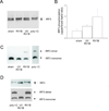
A. BEAS-2B cells were infected with RV1B or UV-irradiated RV1B (1 h at 33°C) at MOI=10. Cell lysates were collected and probed with anti-IRF3 antibody. Poly I:C served as a positive control. IRF3 protein is visualized as two bands, an upper phosphorylated form and a lower unphosphorylated form. B. Densitometry of IRF3 phosphorylation is provided. Bars represent mean ± SEM for 3 experiments. C. Cellular proteins were also subjected to native-PAGE to resolve the dimerization of IRF3. Poly I:C served as a positive control. IRF protein is visualized as two bands, an upper dimer and a lower monomer. (The blot shown is a representative of five individual experiments.) D. Primary tracheobronchial epithelial cells were infected with RV1B or UV-irradiated RV1B. Upper panel: cell lysates were collected twelve h after infection and probed with anti-IRF3 antibody. Poly I:C served as a positive control. Lower panel: cellular proteins were subjected to native-PAGE to resolve the dimerization of IRF3. Poly I:C served as a positive control. (The blots shown are representative of three experiments.)
Figure 11
Figure 11. siRNA against IRF3 blocks RV-induced IFN and ISG expression
A. IRF3 siRNA or non-targeting siRNA was transfected into BEAS-2B while seeding. After transfection, cells were infected with RV1B, UV-irradiated RV1B (UV-RV1B) or sham. A. After infection, cell lysates were probed with anti-IRF3 antibody. (The blot shown is representative of 3 separate experiments.) B–H. Total RNA was extracted and the expression of IFN-β, IFN-λ1, IFN-λ2/3, IRF7, IP-10, IL-8 and GM-CSF was determined by qPCR. The expression of each target gene was normalized to GAPDH. Expression levels are represented as the fold increase vs. sham-infected, non-targeting siRNA-transfected cells. The y-axis has been broken in order to show the effects of siRNA on both basal and maximal gene expression. Bars represent mean ± SEM for four experiments; numbers on top of the bars indicate the fold increase compared to the sham-infected sample within its own siRNA group. (*p<0.05 vs. non-targeting siRNA-transfected RV1B-infected cells, one-way ANOVA.)
Figure 12
Figure 12. siRNA against TRIF, MDA5, but not RIG-I reduced RV1B-induced IRF3 dimerization
A. RIG-I, MDA5, TRIF siRNA or non-targeting siRNA was transfected into BEAS-2B. After transfection, cells were infected with RV1B, UV-irradiated RV1B or sham. After infection, cell lysates were probed with anti-IRF3 antibody. Poly I:C served as a positive control. B. IRF3 dimer to monomer ratio was quantified by densitometry. (The blot shown is a representative of two experiments.)

Similar articles

Cited by

References

    1. Hershenson MB, Johnston SL. Rhinovirus infections: More than a common cold. Am. J. Respir. Crit. Care Med. 2006;174:1284–1285. - PubMed
    1. Yamamoto M, Sato S, Hemmi H, Hoshino K, Kaisho T, Sanjo H, Takeuchi O, Sugiyama M, Okabe M, Takeda K, Akira S. Role of adaptor TRIF in the MyD88-independent Toll-like receptor signaling pathway. Science. 2003;301:640–643. - PubMed
    1. Yoneyama M, Kikuchi M, Natsukawa T, Shinobu N, Imaizumi T, Miyagishi M, Taira K, Akira S, Fujita T. The RNA helicase RIG-I has an essential function in double-stranded RNA-induced innate antiviral responses. Nat. Immunol. 2004;5:730–737. - PubMed
    1. Andrejeva J, Childs KS, Young DF, Carlos TS, Stock N, Goodbourn S, Randall RE. The V proteins of paramyxoviruses bind the IFN-inducible RNA helicase, MDA-5, and inhibit its activation of the IFN-b promoter. Proc. Natl. Acad. Sci. USA. 2004;101:17264–17269. - PMC - PubMed
    1. Kawai T, Takahashi K, Sato S, Coban C, Kumar H, Kato H, Ishii KJ, Takeuchi O, Akira S. IPS-1, an adaptor triggering RIG-I- and MDA5-mediated type I interferon induction. Nat. Immunol. 2005;6:981–988. - PubMed

Publication types

MeSH terms