. 2009 Oct 28;29(43):13543-56.
doi: 10.1523/JNEUROSCI.4144-09.2009.

Simvastatin inhibits the activation of p21ras and prevents the loss of dopaminergic neurons in a mouse model of Parkinson's disease

Affiliations

Simvastatin inhibits the activation of p21ras and prevents the loss of dopaminergic neurons in a mouse model of Parkinson's disease

Anamitra Ghosh et al. J Neurosci. .

Abstract

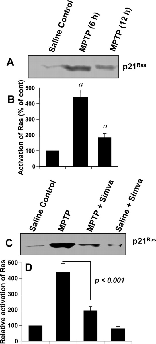
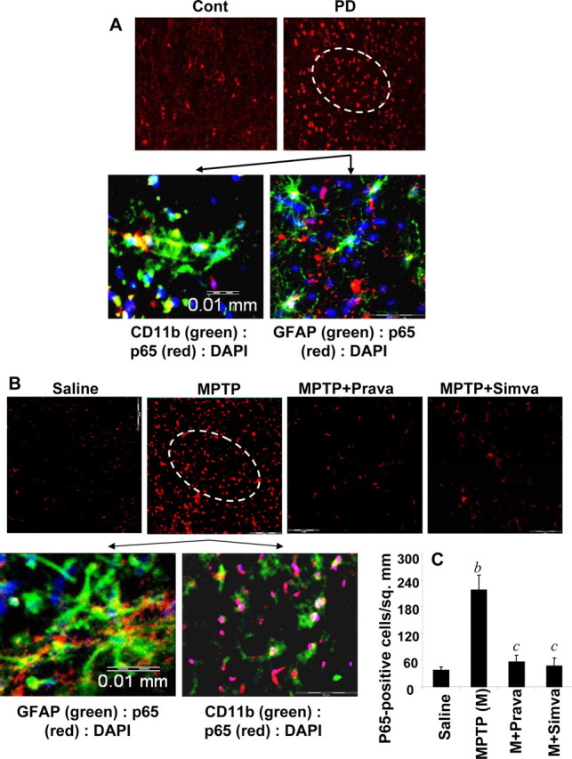
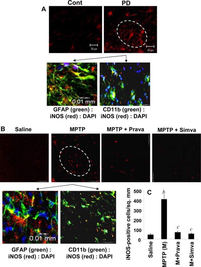
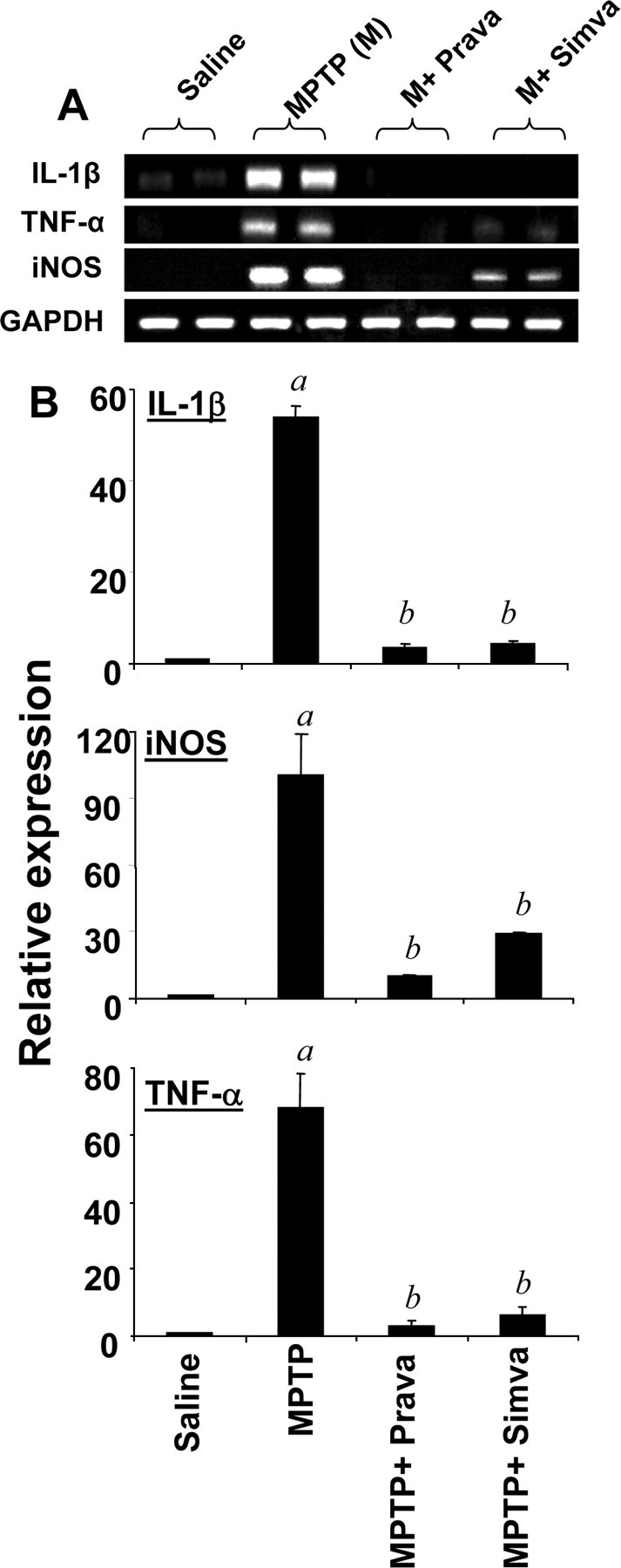
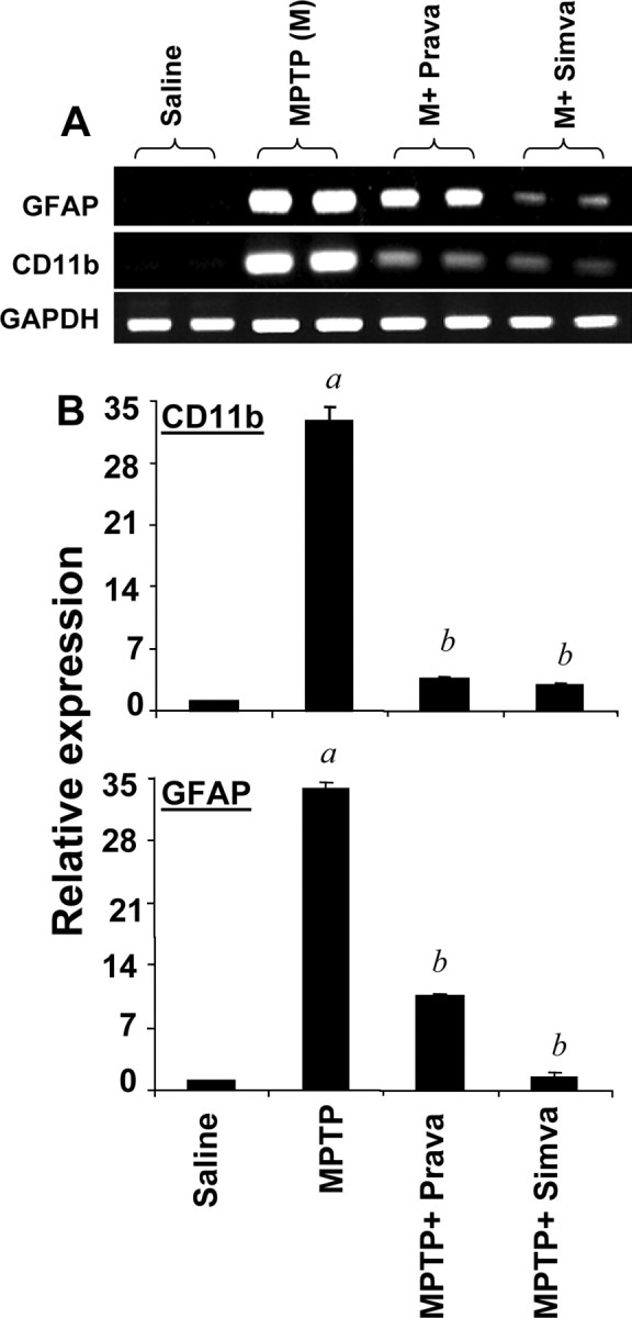
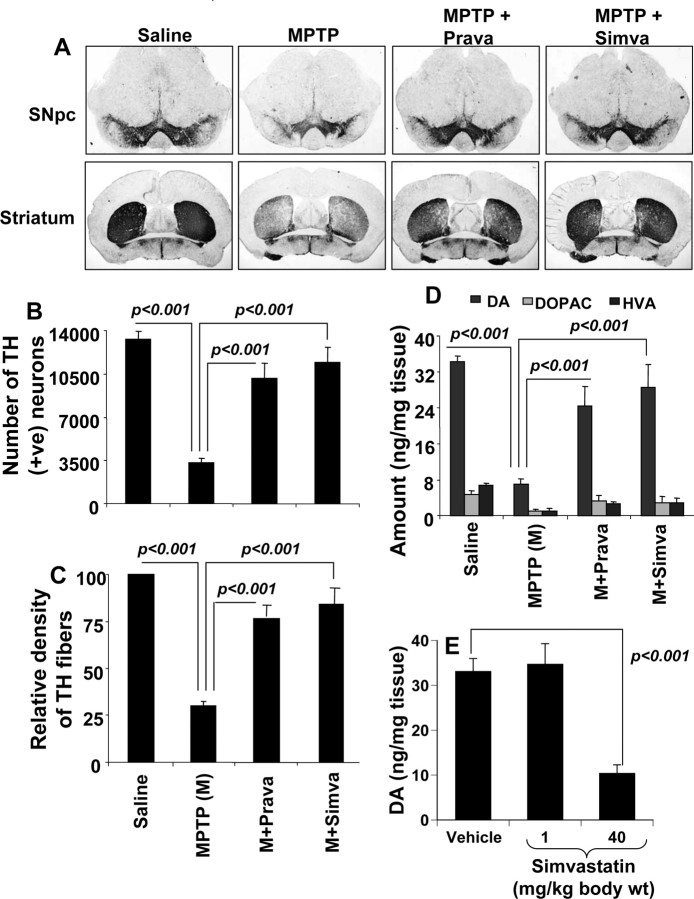
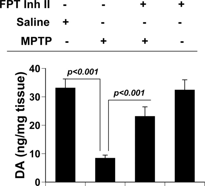
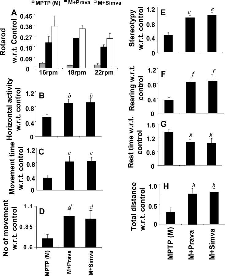
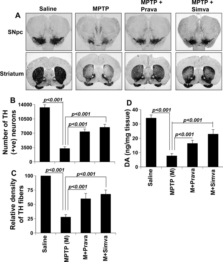
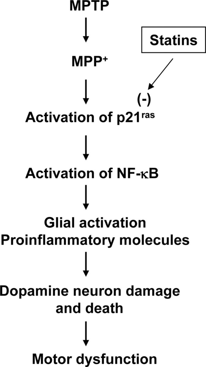
Parkinson's disease (PD) is second only to Alzheimer's disease as the most common devastating human neurodegenerative disorder. Despite intense investigation, no interdictive therapy is available for PD. We investigated whether simvastatin, a Food and Drug Administration-approved cholesterol-lowering drug, could protect against nigrostriatal degeneration after 1-methyl-4-phenyl-1,2,3,6-tetrahydropyridine (MPTP) intoxication to model PD in mice. First, MPP(+) induced the activation of p21(ras) and nuclear factor-kappaB (NF-kappaB) in mouse microglial cells. Inhibition of MPP(+)-induced activation of NF-kappaB by Deltap21(ras), a dominant-negative mutant of p21(ras), supported the involvement of p21(ras) in MPP(+)-induced microglial activation of NF-kappaB. Interestingly, simvastatin attenuated activation of both p21(ras) and NF-kappaB in MPP(+)-stimulated microglial cells. Consistently, we found a very rapid activation of p21(ras) in vivo in the substantia nigra pars compacta of MPTP-intoxicated mice. However, after oral administration, simvastatin entered into the nigra, reduced nigral activation of p21(ras), attenuated nigral activation of NF-kappaB, inhibited nigral expression of proinflammatory molecules, and suppressed nigral activation of glial cells. These findings paralleled dopaminergic neuronal protection, normalized striatal neurotransmitters, and improved motor functions in MPTP-intoxicated mice. Similarly, pravastatin, another cholesterol-lowering drug, suppressed microglial inflammatory responses and protected dopaminergic neurons in MPTP-intoxicated mice, but at levels less than simvastatin. Furthermore, both the statins administered 2 d after initiation of the disease were still capable of inhibiting the demise of dopaminergic neurons and concomitant loss of neurotransmitters, suggesting that statins are capable of slowing down the progression of neuronal loss in the MPTP mouse model. Therefore, we conclude that statins may be of therapeutic benefit for PD patients.


Figures

Figure 1.
Figure 1.
MPP+ induces the activation of p21ras and NF-κB in mouse BV-2 microglial cells via simvastatin-sensitive pathway. A, Cells were stimulated with 1 μm MPP+ under serum-free condition. At different time points, activation of p21ras was monitored (top panel) as described in Materials and Methods. In another set, cells pretreated with 10 μm simvastatin for 6 h were challenged with 1 μm MPP+ under serum-free condition followed by monitoring activation of p21ras at different time points (bottom panel). B, Bands from three different experiments were quantified and activation of Ras is shown as percentage of control. ap < 0.0001 versus control (0 min). C, Cells were cotransfected with 0.25 μg of pBIIX-Luc and 12.5 ng of pRL-TK. After 24 h of transfection, cells were stimulated with different concentrations of MPP+ under serum-free condition. After 6 h of stimulation, firefly and Renilla luciferase activities were obtained by analyzing total cell extract. Data are mean + SEM of three separate experiments. bp < 0.0001 versus control (0 μm). D, Cells were cotransfected with 0.125 μg of pBIIX-Luc, 0.125 μg of either an empty vector or Δp21ras, and 12.5 ng of pRL-TK. After 24 h of transfection, cells were stimulated with MPP+ under serum-free condition. After 6 h of stimulation, firefly and Renilla luciferase activities were analyzed. E, Cells were cotransfected with 0.25 μg of pBIIX-Luc and 12.5 ng of pRL-TK. After 24 h of transfection, cells were treated with different concentrations of simvastatin for 6 h followed by stimulation with MPP+. After 6 h of stimulation, firefly and Renilla luciferase activities were analyzed. Data are mean + SD of three separate experiments. cp < 0.001 versus MPP+.
Figure 2.
Figure 2.
Activation of p21ras in ventral midbrain of MPTP-intoxicated mice is simvastatin sensitive. A, Six and 12 h after the last injection of MPTP, activation of p21ras was monitored in ventral midbrain tissues. Saline was used as control. The experiment was repeated four times, each time on one animal in each group. B, Bands from four different mice were quantified, and activation of Ras is shown as percentage of control. ap < 0.001 versus saline control. C, Mice were treated with simvastatin (1 mg · kg body weight−1 · d−1) via gavage from 24 h before MPTP injection. Six hours after the last injection of MPTP, activation of p21ras was monitored in ventral midbrain tissues. The experiment was repeated four times each time using one animal in each group. D, Bands from four different mice were quantified and activation of p21ras is shown as percentage of control. Error bars indicate SEM.
Figure 3.
Figure 3.
Activation of NF-κB in ventral midbrain of MPTP-intoxicated mice is statin sensitive. A, Midbrain sections of PD patients and age-matched controls were immunostained for p65 (low magnification). Sections of PD patients were also double-labeled for p65 and glial cell markers (GFAP and CD11b). Results represent three independent experiments. B, Mice were treated with pravastatin and simvastatin (1 mg · kg body weight−1 · d−1) from 3 h after the last injection of MPTP. Twenty-four hours after the last injection of MPTP, ventral midbrain sections were immunostained for p65 (low magnification). Midbrain sections of MPTP-intoxicated mice were also double-labeled for p65 and glial cell markers (GFAP for astrocytes and CD11b for microglia). Results represent three independent experiments. C, NF-κB p65-positive cells were counted in four nigral sections (2 images per slide) from each of four mice. bp < 0.0001 versus saline-control; cp < 0.0001 versus MPTP. Error bars indicate SEM.
Figure 4.
Figure 4.
Entry of statins into the nigra of MPTP-intoxicated mice. Mice were treated with pravastatin and simvastatin (1 mg · kg body weight−1 · d−1) from 24 h before MPTP injection. Twenty-four hours after the last injection of MPTP, nigra was dissected out and concentrations of pravastatin and simvastatin were analyzed in nigral samples by HPLC using ibuprofen as an internal standard. A, MPTP. B, MPTP plus pravastatin. C, MPTP plus simvastatin.
Figure 5.
Figure 5.
Expression of iNOS in ventral midbrain of MPTP-intoxicated mice is statin sensitive. A, Midbrain sections of PD patients and age-matched controls were immunostained for iNOS (low magnification). Sections of PD patients were also double-labeled for iNOS and glial cell markers (GFAP and CD11b). Results represent three independent experiments. B, Mice were treated with pravastatin and simvastatin (1 mg · kg body weight−1 · d−1) from 3 h after the last injection of MPTP. Twenty-four hours after the last injection of MPTP, ventral midbrain sections were immunostained for iNOS (low magnification). Midbrain sections of MPTP-intoxicated mice were also double-labeled for iNOS and glial cell markers (GFAP for astrocytes and CD11b for microglia). Results represent three independent experiments. C, Cells positive for iNOS were counted in four nigral sections (2 images per slide) from each of four mice. bp < 0.0001 versus saline-control; cp < 0.0001 versus MPTP. Error bars indicate SEM.
Figure 6.
Figure 6.
Simvastatin and pravastatin inhibit the expression of iNOS and proinflammatory cytokines in ventral midbrain of MPTP-intoxicated mice. Mice receiving simvastatin and pravastatin (1 mg · kg body weight−1 · d−1) from 3 h after the last injection of MPTP were killed 24 h after the last injection of MPTP. The mRNA expression of iNOS, IL-1β, and TNF-α was analyzed by semiquantitative RT-PCR (A) and quantitative real-time PCR (B). Data are means + SEM of five mice per group. ap < 0.0001 versus saline group; bp < 0.0001 versus the MPTP group.
Figure 7.
Figure 7.
Increased expression of CD11b and GFAP in ventral midbrain of MPTP-intoxicated mice is statin sensitive. A, Midbrain sections of PD patients and age-matched controls were immunostained for GFAP and CD11b. B, Mice were treated with pravastatin and simvastatin (1 mg · kg body weight−1 · d−1) from 3 h after the last injection of MPTP. Twenty-four hours after the last injection of MPTP, ventral midbrain sections were immunostained for GFAP and CD11b. Results represent analysis of four nigral sections (2 images per slide) from each of four mice.
Figure 8.
Figure 8.
Simvastatin and pravastatin inhibit mRNA expression of GFAP and CD11b in ventral midbrain of MPTP-intoxicated mice. Mice receiving simvastatin and pravastatin (1 mg · kg body weight−1 · d−1) from 3 h after the last injection of MPTP were killed 24 h after the last injection of MPTP. The mRNA expression of GFAP and CD11b was analyzed by semiquantitative RT-PCR (A) and quantitative real-time PCR (B). Data are means + SEM of five mice per group. ap < 0.0001 versus saline group; bp < 0.0001 versus the MPTP group.
Figure 9.
Figure 9.
Simvastatin and pravastatin protect dopaminergic neurons in MPTP-intoxicated mice. Mice receiving simvastatin and pravastatin (1 mg · kg body weight−1 · d−1) from 3 h after the last injection of MPTP were killed 7 d after the last injection of MPTP followed by TH immunostaining of SNpc (top panel) and striatum (bottom panel) (A), counting of TH-positive neurons in SNpc (B), quantification of TH-positive fibers in striatum (C), and assay of neurotransmitters in striatum (D). To understand whether statin alone was toxic for the nigrostriatum, groups of mice received two different doses of simvastatin (1 and 40 mg · kg body weight−1 · d−1). After 7 d, the concentration of dopamine was assayed in striatum (E). Data are means + SEM of eight mice per group.
Figure 10.
Figure 10.
Effect of FPT inhibitor II on the loss of dopamine in MPTP-intoxicated mice. Mice receiving FPT inhibitor II (10 mg · kg body weight−1 · d−1) from 3 h after the last injection of MPTP were killed 7 d after the last injection of MPTP followed by assay of dopamine in striatum (DA). Data are means + SEM of eight mice per group.
Figure 11.
Figure 11.
Simvastatin and pravastatin improve motor functions in MPTP-intoxicated mice. Mice receiving simvastatin and pravastatin (1 mg · kg body weight−1 · d−1) from 3 h after the last injection of MPTP were tested for motor functions [rotorod (A); horizontal activity (B); movement time (C); number of movements (D); stereotypy counts (E); rearing (F); rest time (G); total distance (H)] 7 d after the last injection of MPTP. Data are means + SEM of eight mice per group. b,c,e,f,hp < 0.001 versus MPTP. d,gp < 0.05 versus MPTP. For rotorod, we performed two-way ANOVA analysis and observed significant improvement in rotorod performance by simvastatin and pravastatin at 16 rpm (F(2,21) = 72.35; p < 0.0001), 18 rpm (F(2,21) = 170.41; p < 0.0001), and 22 rpm (F(2,21) = 108.94; p < 0.0001). Error bars indicate SEM.
Figure 12.
Figure 12.
Simvastatin and pravastatin suppress disease progression in the MPTP mouse model. Mice receiving simvastatin and pravastatin (1 mg · kg body weight−1 · d−1) from 2 d after the last injection of MPTP were killed 7 d after the last injection of MPTP followed by TH immunostaining of SNpc (top panel) and striatum (bottom panel) (A), counting of TH-positive neurons in SNpc (B), quantification of TH-positive fibers in striatum (C), and assay of neurotransmitters in striatum (D). Data are means + SEM of eight mice per group.
Figure 13.
Figure 13.
Schematic presentation of possible hypothesis.

Similar articles

Cited by

References

    1. Benner EJ, Mosley RL, Destache CJ, Lewis TB, Jackson-Lewis V, Gorantla S, Nemachek C, Green SR, Przedborski S, Gendelman HE. Therapeutic immunization protects dopaminergic neurons in a mouse model of Parkinson's disease. Proc Natl Acad Sci U S A. 2004;101:9435–9440. - PMC - PubMed
    1. Bessler H, Djaldetti R, Salman H, Bergman M, Djaldetti M. IL-1 beta, IL-2, IL-6 and TNF-alpha production by peripheral blood mononuclear cells from patients with Parkinson's disease. Biomed Pharmacother. 1999;53:141–145. - PubMed
    1. Brahmachari S, Fung YK, Pahan K. Induction of glial fibrillary acidic protein expression in astrocytes by nitric oxide. J Neurosci. 2006;26:4930–4939. - PMC - PubMed
    1. Brochard V, Combadière B, Prigent A, Laouar Y, Perrin A, Beray-Berthat V, Bonduelle O, Alvarez-Fischer D, Callebert J, Launay JM, Duyckaerts C, Flavell RA, Hirsch EC, Hunot S. Infiltration of CD4+ lymphocytes into the brain contributes to neurodegeneration in a mouse model of Parkinson disease. J Clin Invest. 2009;119:182–192. - PMC - PubMed
    1. Dasgupta S, Jana M, Liu X, Pahan K. Role of very-late antigen-4 (VLA-4) in myelin basic protein-primed T cell contact-induced expression of proinflammatory cytokines in microglial cells. J Biol Chem. 2003;278:22424–22431. - PMC - PubMed

Publication types

MeSH terms