Selective and specific macrophage ablation is detrimental to wound healing in mice
- PMID: 19850888
- PMCID: PMC2789630
- DOI: 10.2353/ajpath.2009.090248
Selective and specific macrophage ablation is detrimental to wound healing in mice
Abstract
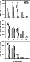
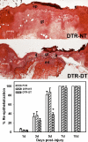
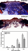
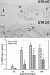
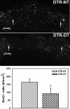
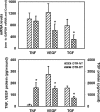
Macrophages are thought to play important roles during wound healing, but definition of these roles has been hampered by our technical inability to specifically eliminate macrophages during wound repair. The purpose of this study was to test the hypothesis that specific depletion of macrophages after excisional skin wounding would detrimentally affect healing by reducing the production of growth factors important in the repair process. We used transgenic mice that express the human diphtheria toxin (DT) receptor under the control of the CD11b promoter (DTR mice) to specifically ablate macrophages during wound healing. Mice without the transgene are relatively insensitive to DT, and administration of DT to wild-type mice does not alter macrophage or other inflammatory cell accumulation after injury and does not influence wound healing. In contrast, treatment of DTR mice with DT prevented macrophage accumulation in healing wounds but did not affect the accumulation of neutrophils or monocytes. Such macrophage depletion resulted in delayed re-epithelialization, reduced collagen deposition, impaired angiogenesis, and decreased cell proliferation in the healing wounds. These adverse changes were associated with increased levels of tumor necrosis factor-alpha and reduced levels of transforming growth factor-beta1 and vascular endothelial growth factor in the wound. In summary, macrophages seem to promote both wound closure and dermal healing, in part by regulating the cytokine environment of the healing wound.
Figures
Similar articles
-
A transgenic mouse model of inducible macrophage depletion: effects of diphtheria toxin-driven lysozyme M-specific cell lineage ablation on wound inflammatory, angiogenic, and contractive processes.Am J Pathol. 2009 Jul;175(1):132-47. doi: 10.2353/ajpath.2009.081002. Epub 2009 Jun 15. Am J Pathol. 2009. PMID: 19528348 Free PMC article.
-
Macrophages are essential contributors to kidney injury in murine cryoglobulinemic membranoproliferative glomerulonephritis.Kidney Int. 2011 Nov;80(9):946-958. doi: 10.1038/ki.2011.249. Epub 2011 Aug 3. Kidney Int. 2011. PMID: 21814168
-
TNF-alpha-dependent regulation of acute pancreatitis severity by Ly-6C(hi) monocytes in mice.J Biol Chem. 2011 Apr 15;286(15):13327-35. doi: 10.1074/jbc.M111.218388. Epub 2011 Feb 22. J Biol Chem. 2011. PMID: 21343291 Free PMC article.
-
Role of macrophages in normal wound healing: an overview.J Wound Care. 2009 Aug;18(8):349-51. doi: 10.12968/jowc.2009.18.8.43636. J Wound Care. 2009. PMID: 19862875 Review.
-
Wound macrophages as key regulators of repair: origin, phenotype, and function.Am J Pathol. 2011 Jan;178(1):19-25. doi: 10.1016/j.ajpath.2010.08.003. Epub 2010 Dec 23. Am J Pathol. 2011. PMID: 21224038 Free PMC article. Review.
Cited by
-
The Immune System in Transfusion-Related Acute Lung Injury Prevention and Therapy: Update and Perspective.Front Mol Biosci. 2021 Mar 24;8:639976. doi: 10.3389/fmolb.2021.639976. eCollection 2021. Front Mol Biosci. 2021. PMID: 33842545 Free PMC article. Review.
-
Mesenchymal stromal exosome-functionalized scaffolds induce innate and adaptive immunomodulatory responses toward tissue repair.Sci Adv. 2021 May 12;7(20):eabf7207. doi: 10.1126/sciadv.abf7207. Print 2021 May. Sci Adv. 2021. PMID: 33980490 Free PMC article.
-
Chronic wound repair and healing in older adults: current status and future research.J Am Geriatr Soc. 2015 Mar;63(3):427-38. doi: 10.1111/jgs.13332. Epub 2015 Mar 6. J Am Geriatr Soc. 2015. PMID: 25753048 Free PMC article.
-
Transition from inflammation to proliferation: a critical step during wound healing.Cell Mol Life Sci. 2016 Oct;73(20):3861-85. doi: 10.1007/s00018-016-2268-0. Epub 2016 May 14. Cell Mol Life Sci. 2016. PMID: 27180275 Free PMC article. Review.
-
H-ferritin and CD68(+) /H-ferritin(+) monocytes/macrophages are increased in the skin of adult-onset Still's disease patients and correlate with the multi-visceral involvement of the disease.Clin Exp Immunol. 2016 Oct;186(1):30-8. doi: 10.1111/cei.12826. Epub 2016 Jul 28. Clin Exp Immunol. 2016. PMID: 27317930 Free PMC article.
References
-
- Eming SA, Krieg T, Davidson JM. Inflammation in wound repair: molecular and cellular mechanisms. J Invest Dermatol. 2007;127:514–525 17299434. - PubMed
-
- Singer AJ, Clark RA. Cutaneous wound healing. N Engl J Med. 1999;341:738–746. - PubMed
-
- Martin P. Wound healing—aiming for perfect skin regeneration. Science. 1997;276:75–81. - PubMed
-
- Loots MA, Lamme EN, Zeegelaar J, Mekkes JR, Bos JD, Middelkoop E. Differences in cellular infiltrate and extracellular matrix of chronic diabetic and venous ulcers versus acute wounds. J Invest Dermatol. 1998;111:850–857. - PubMed
Publication types
MeSH terms
Substances
Grants and funding
LinkOut - more resources
Full Text Sources
Other Literature Sources
Molecular Biology Databases
Research Materials

