

Diabetes regulates mitochondrial biogenesis and fission in mouse neurons
- PMID: 19847394
- PMCID: PMC4011390
- DOI: 10.1007/s00125-009-1553-y
Diabetes regulates mitochondrial biogenesis and fission in mouse neurons
Abstract
Aims/hypothesis: Normal mitochondrial activity is a critical component of neuronal metabolism and function. Disruption of mitochondrial activity by altered mitochondrial fission and fusion is the root cause of both neurodegenerative disorders and Charcot-Marie-Tooth type 2A inherited neuropathy. This study addressed the role of mitochondrial fission in the pathogenesis of diabetic neuropathy.
Methods: Mitochondrial biogenesis and fission were assayed in both in vivo and in vitro models of diabetic neuropathy. Gene, protein, mitochondrial DNA and ultrastructural analyses were used to assess mitochondrial biogenesis and fission.
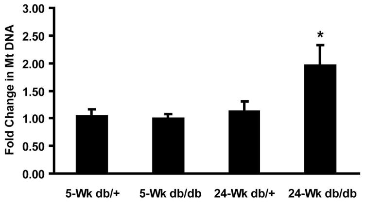
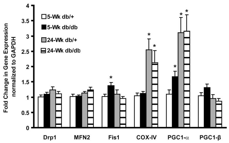
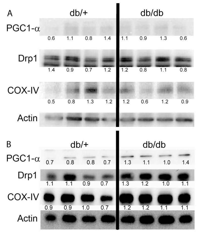
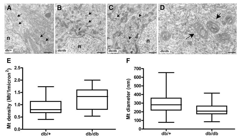
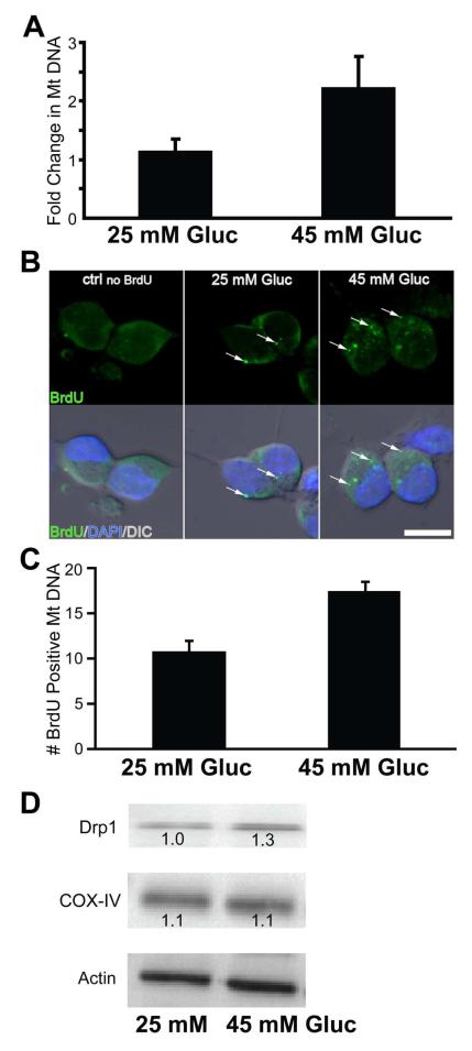
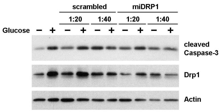
Results: There was greater mitochondrial biogenesis in dorsal root ganglion neurons from diabetic compared with non-diabetic mice. An essential step in mitochondrial biogenesis is mitochondrial fission, regulated by the mitochondrial fission protein dynamin-related protein 1 (DRP1). Evaluation of diabetic neurons in vivo indicated small, fragmented mitochondria, suggesting increased fission. In vitro studies revealed that short-term hyperglycaemic exposure increased levels of DRP1 protein. The influence of hyperglycaemia-mediated mitochondrial fission on cell viability was evaluated by knockdown of Drp1 (also known as Dnm1l). Knockdown of Drp1 resulted in decreased susceptibility to hyperglycaemic damage.
Conclusions/interpretation: We propose that: (1) mitochondria undergo biogenesis in response to hyperglycaemia, but the increased biogenesis is insufficient to accommodate the metabolic load; (2) hyperglycaemia causes an excess of mitochondrial fission, creating small, damaged mitochondria; and (3) reduction of aberrant mitochondrial fission increases neuronal survival and indicates an important role for the fission-fusion equilibrium in the pathogenesis of diabetic neuropathy.
Figures
Similar articles
-
Mitochondria in DRG neurons undergo hyperglycemic mediated injury through Bim, Bax and the fission protein Drp1.Neurobiol Dis. 2006 Jul;23(1):11-22. doi: 10.1016/j.nbd.2006.01.017. Epub 2006 May 8. Neurobiol Dis. 2006. PMID: 16684605
-
Mitochondrial biogenesis and fission in axons in cell culture and animal models of diabetic neuropathy.Acta Neuropathol. 2010 Oct;120(4):477-89. doi: 10.1007/s00401-010-0697-7. Epub 2010 May 15. Acta Neuropathol. 2010. PMID: 20473509 Free PMC article.
-
PGC-1α regulation of mitochondrial degeneration in experimental diabetic neuropathy.Neurobiol Dis. 2014 Apr;64:118-30. doi: 10.1016/j.nbd.2014.01.001. Epub 2014 Jan 11. Neurobiol Dis. 2014. PMID: 24423644 Free PMC article.
-
The Drp1-Mediated Mitochondrial Fission Protein Interactome as an Emerging Core Player in Mitochondrial Dynamics and Cardiovascular Disease Therapy.Int J Mol Sci. 2023 Mar 17;24(6):5785. doi: 10.3390/ijms24065785. Int J Mol Sci. 2023. PMID: 36982862 Free PMC article. Review.
-
Dynamin-related protein 1 and mitochondrial fragmentation in neurodegenerative diseases.Brain Res Rev. 2011 Jun 24;67(1-2):103-18. doi: 10.1016/j.brainresrev.2010.11.004. Epub 2010 Dec 8. Brain Res Rev. 2011. PMID: 21145355 Free PMC article. Review.
Cited by
-
pS421 huntingtin modulates mitochondrial phenotypes and confers neuroprotection in an HD hiPSC model.Cell Death Dis. 2020 Sep 25;11(9):809. doi: 10.1038/s41419-020-02983-z. Cell Death Dis. 2020. PMID: 32978366 Free PMC article.
-
Modulating mitochondrial fission to lower diabetic oxidative stress.Diabetes. 2012 Aug;61(8):1915-7. doi: 10.2337/db12-0463. Diabetes. 2012. PMID: 22826308 Free PMC article. No abstract available.
-
Visualization of mitochondrial DNA replication in individual cells by EdU signal amplification.J Vis Exp. 2010 Nov 15;(45):2147. doi: 10.3791/2147. J Vis Exp. 2010. PMID: 21113116 Free PMC article.
-
Impairment of Novel Object Recognition Memory and Brain Insulin Signaling in Fructose- but Not Glucose-Drinking Female Rats.Mol Neurobiol. 2018 Aug;55(8):6984-6999. doi: 10.1007/s12035-017-0863-1. Epub 2018 Jan 26. Mol Neurobiol. 2018. PMID: 29372547
-
What Is in the Field for Genetics and Epigenetics of Diabetic Neuropathy: The Role of MicroRNAs.J Diabetes Res. 2021 Oct 6;2021:5593608. doi: 10.1155/2021/5593608. eCollection 2021. J Diabetes Res. 2021. PMID: 34660810 Free PMC article. Review.
References
-
- Leinninger GM, Backus C, Sastry AM, Yi YB, Wang CW, Feldman EL. Mitochondria in DRG neurons undergo hyperglycemic mediated injury through Bim, Bax and the fission protein Drp1. Neurobiology of disease. 2006;23:11–22. - PubMed
-
- Cartoni R, Martinou JC. Role of mitofusin 2 mutations in the physiopathology of Charcot-Marie-Tooth disease type 2A. Exp Neurol. 2009;218:268–273. - PubMed
-
- Vincent AM, McLean LL, Backus C, Feldman EL. Short-term hyperglycemia produces oxidative damage and apoptosis in neurons. FASEB J. 2005;19:638–640. - PubMed
-
- Huang TJ, Price SA, Chilton L, et al. Insulin prevents depolarization of the mitochondrial inner membrane in sensory neurons of type 1 diabetic rats in the presence of sustained hyperglycemia. Diabetes. 2003;52:2129–2136. - PubMed
Publication types
MeSH terms
Substances
Grants and funding
LinkOut - more resources
Full Text Sources
Molecular Biology Databases
Research Materials
Miscellaneous
