. 2010 Jan;84(1):280-90.
doi: 10.1128/JVI.01772-09.

Murine hepatitis virus nonstructural protein 4 regulates virus-induced membrane modifications and replication complex function

Affiliations

Murine hepatitis virus nonstructural protein 4 regulates virus-induced membrane modifications and replication complex function

Mark J Gadlage et al. J Virol. 2010 Jan.

Abstract

Positive-strand RNA viruses induce modifications of cytoplasmic membranes to form replication complexes. For coronaviruses, replicase nonstructural protein 4 (nsp4) has been proposed to function in the formation and organization of replication complexes. Murine hepatitis virus (MHV) nsp4 is glycosylated at residues Asn176 (N176) and N237 during plasmid expression of nsp4 in cells. To test if MHV nsp4 residues N176 and N237 are glycosylated during virus replication and to determine the effects of N176 and N237 on nsp4 function and MHV replication, alanine substitutions of nsp4 N176, N237, or both were engineered into the MHV-A59 genome. The N176A, N237A, and N176A/N237A mutant viruses were viable, and N176 and N237 were glycosylated during infection of wild-type (wt) and mutant viruses. The nsp4 glycosylation mutants exhibited impaired virus growth and RNA synthesis, with the N237A and N176A/N237A mutant viruses demonstrating more profound defects in virus growth and RNA synthesis. Electron microscopic analysis of ultrastructure from infected cells demonstrated that the nsp4 mutants had aberrant morphology of virus-induced double-membrane vesicles (DMVs) compared to those infected with wt virus. The degree of altered DMV morphology directly correlated with the extent of impairment in viral RNA synthesis and virus growth of the nsp4 mutant viruses. The results indicate that nsp4 plays a critical role in the organization and stability of DMVs. The results also support the conclusion that the structure of DMVs is essential for efficient RNA synthesis and optimal replication of coronaviruses.


Figures

FIG. 1.
FIG. 1.
Processing, glycosylation, and mutagenesis of nsp4. (A) Schematic of MHV nsp4 processing. Three virus-encoded proteases process pp1ab into intermediate precursors and 16 mature nsp's. PLP1 and PLP2 are shown as black boxes within nsp3, while the nsp5 protease (3CLpro) is shown in gray. PLP-mediated processing of nsp's is linked by white boxes, and 3CLpro processing is linked by gray boxes. Nsp4 is shown in black. Nsp's are indicated by number. The nsp4-to-10 precursor is also shown. (B) Proposed topology and N-linked glycosylation sites of nsp4. MHV nsp4 is a 496-amino acid protein that has four predicted transmembrane domains (TM1 to 4, black rectangles) and five soluble regions (SRa to e). Locations of N-linked glycosylation residues Asn176 and Asn237 (N176 and N237) are indicated in SRb, and predicted luminal and cytoplasmic domains are indicated (35). (C) Engineered nsp4 glycosylation mutants. Nsp4 glycosylation mutants were engineered by replacing the AAT asparagine codons at both N176 and N237 with a GCC alanine codon. Nucleotide numbers correspond to genomic position, and amino acid numbers correspond to nsp4 position.
FIG. 2.
FIG. 2.
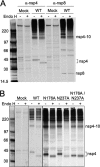
Protein expression and glycosylation of nsp4. Cytoplasmic lysates were generated from radiolabeled DBT cells that were either mock infected or infected with wt, N176A, N237A, or N176A/N237A viruses. Labeled proteins were immunoprecipitated using antiserum against nsp4 or nsp8. (A) Endo H treatment of wt nsp4 and nsp8. Immunoprecipitated nsp4 and nsp8 were either mock treated or treated with Endo H to analyze N-linked glycosylation. After Endo H treatment for 3 h, proteins were resolved on SDS-PAGE and visualized by fluorography. Black dots indicate either glycosylated or unglycosylated forms of nsp4. α-nsp4, anti-nsp4; α-nsp8, anti-nsp8. (B) Endo H treatment of nsp4 glycosylation mutants. Immunoprecipitated nsp4 from the wt or nsp4 glycosylation mutants was mock treated or treated with Endo H. All samples in each panel were resolved on the same gel and had the same exposure time, but the images shown in panel B were cropped to remove nonrelevant lanes. Molecular weight markers (in thousands) are shown to the left of each gel.
FIG. 3.
FIG. 3.
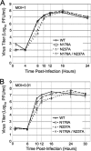
Growth analysis of nsp4 glycosylation mutant viruses. DBT cells were infected with the indicated viruses for single cycle growth at an MOI of 1 PFU/cell for 24 h (A) or for multiple cycle growth at an MOI of 0.01 PFU/cell for 30 h (B). Samples of virus supernatants were collected at the times indicated beneath the graphs. Virus titers were determined by plaque assay with DBT cells. Error bars represent standard deviations from the mean based on samples from multiple replicates.
FIG. 4.
FIG. 4.
RNA synthesis of nsp4 glycosylation mutant viruses. DBT cells in six-well plates were mock infected or infected with wt, N176A, N237A, or N176A/N237A viruses at an MOI of 5 PFU/cell. Cells were treated with Act D for 30 min prior to addition of radiolabel. Cells were metabolically labeled with [3H]uridine for the intervals indicated, cells were lysed, and [3H]uridine incorporation was quantified by liquid scintillation counting of TCA-precipitable RNA. Data points represent the mean counts/minute (CPM) of two individual experiments, and error bars represent the standard deviations between two experiments.
FIG. 5.
FIG. 5.
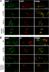
Immunofluorescence of nsp4 localization. DBT cells on glass coverslips were infected with the indicated viruses at an MOI of 10 PFU/cell. At 6 h p.i., cells were fixed, probed with antibodies to nsp4, nsp8, and membrane (M) protein, and analyzed by immunofluorescence using a Zeiss Axiovert 200 microscope at ×40 magnification. (A) nsp4 colocalizes with nsp8. Infected cells were analyzed by indirect immunofluorescence using anti-nsp4 (Alexa 488, green) and direct immunofluorescence by Alexa 546 conjugated to anti-nsp8 (red). Yellow pixels represent colocalization of overlapping green and red pixels. (B) nsp4 does not colocalize with M protein. Infected cells were probed by indirect immunofluorescence using rabbit anti-nsp4 (green) and mouse anti-M (red). The scale bar in the upper images in panels A and B equals 20 μM and is representative of all other images.
FIG. 6.
FIG. 6.
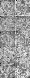
TEM analysis of replication complexes and DMVs from the wt and nsp4 mutants. DBT cells were mock infected or infected with wt, N176A, N237A, or N176A/N237A viruses. Cells were harvested in 2% glutaraldehyde and processed for TEM analysis. (A and A1) Mock-infected cells. (B and B1) wt MHV infection. (C and C1) N176A mutant virus infection. (D and D1) N237A mutant virus infection. (E and E1) N176A/N237A mutant virus infection. Dotted boxes in the left images indicate area of magnification in right image. The scale bar in the left images represents 500 nm. Arrowheads indicate dark-stained, individual virions, which are located above the arrowheads. Black arrows point to CMs. * indicates examples of regular DMV structure. ⊗ shows examples of irregular DMV structure. N, nucleus; G, Golgi apparatus; M, mitochondria.
FIG. 7.
FIG. 7.
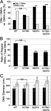
Quantitative analysis of CMs and DMVs. (A) CMs and DMVs. All EM images were analyzed for the presence of CMs and DMVs based on characteristic EM morphology. Because CMs were found only in the presence of DMVs in all TEM sections observed, the ratio of total cell sections with CMs plus DMVs to the total of cell sections with DMVs alone could be examined. Black bars indicate presence of both CMs and DMVs, while white bars represent the presence of DMVs alone. Chi-square analysis was used to compare the presence of CMs plus DMVs to DMVs alone. (B) Ratios of DMVs with regular morphology to total DMVs (regular plus irregular). Total DMVs and DMVs with regular morphology were counted with TEM images for all viruses, and the ratio of regular DMVs to total DMVs was determined. (C) Diameter of regular and irregular DMVs of the wt and nsp4 mutants. DMVs were measured in Image J by the widest diameter in nm of outer membranes. Black bars indicate regular DMVs, while white bars indicate irregular DMVs. Error bars indicate standard deviation. There was no significant difference (not labeled in the figure) in the diameters of regular DMVs between wt and nsp4 mutant viruses. ANOVA followed by Tukey tests indicated a significant difference in the diameters of irregular DMVs of the N237A and N176A/N237A viruses compared to those of both wt and N176A viruses. * (P < 0.05), ** (P < 0.01), and *** (p < 0.001) indicate levels of statistical significance compared to wt virus. NS, no significance.

Similar articles

Cited by

References

    1. Baker, S. C., K. Yokomori, S. Dong, R. Carlisle, A. E. Gorbalenya, E. V. Koonin, and M. M. Lai. 1993. Identification of the catalytic sites of a papain-like cysteine proteinase of murine coronavirus. J. Virol. 67:6056-6063. - PMC - PubMed
    1. Baliji, S., S. A. Cammer, B. Sobral, and S. C. Baker. 2009. Detection of nonstructural protein 6 (nsp6) in murine coronavirus-infected cells and analysis of the transmembrane topology using bioinformatics and molecular approaches. J. Virol. 83:6957-6962. - PMC - PubMed
    1. Bonilla, P. J., A. E. Gorbalenya, and S. R. Weiss. 1994. Mouse hepatitis virus strain A59 RNA polymerase gene ORF 1a: heterogeneity among MHV strains. Virology 198:736-740. - PMC - PubMed
    1. Bost, A. G., E. Prentice, and M. R. Denison. 2001. Mouse hepatitis virus replicase protein complexes are translocated to sites of M protein accumulation in the ERGIC at late times of infection. Virology 285:21-29. - PMC - PubMed
    1. Bredenbeek, P. J., C. J. Pachuk, A. F. H. Noten, J. Charite, W. Luytjes, S. R. Weiss, and W. J. M. Spaan. 1990. The primary structure and expression of the second open reading frame of the polymerase gene of the coronavirus MHV-A59; a highly conserved polymerase is expressed by an efficient ribosomal frameshifting mechanism. Nucleic Acids Res. 18:1825-1832. - PMC - PubMed

LinkOut - more resources