. 2010 Jan 15;19(2):235-49.
doi: 10.1093/hmg/ddp482. Epub 2009 Oct 19.

Proteotoxic stress increases nuclear localization of ataxin-3

Affiliations

Proteotoxic stress increases nuclear localization of ataxin-3

Christopher P Reina et al. Hum Mol Genet. .

Abstract

Spinocerebellar ataxia type 3 (SCA3)/Machado Joseph disease results from expansion of the polyglutamine domain in ataxin-3 (Atx3). Atx3 is a transcriptional co-repressor, as well as a deubiquitinating enzyme that appears to function in cellular pathways involved in protein homeostasis. In this study, we show that interactions of Atx3 with valosin-containing protein and hHR23B are dynamic and modulated by proteotoxic stresses. Heat shock, a general proteotoxic stress, also induced wild-type and pathogenic Atx3 to accumulate in the nucleus. Mapping studies showed that two regions of Atx3, the Josephin domain and the C-terminus, regulated heat shock-induced nuclear localization. Heat shock-induced nuclear localization of Atx3 was not affected by a casein kinase-2 inhibitor or by mutating a predicted nuclear localization signal. However, serine-111 of Atx3 was required for nuclear localization of the Josephin domain and regulated nuclear localization of full-length Atx3. Atx3 null cells were more sensitive to toxic effects of heat shock suggesting that Atx3 had a protective function in the cellular response to heat shock. Importantly, we found that oxidative stress also induced nuclear localization of Atx3; both wild-type and pathogenic Atx3 accumulated in the nucleus of SCA3 patient fibroblasts following oxidative stress. Heat shock and oxidative stress are the first processes identified that increase nuclear localization of Atx3. Observations in this study provide new and important insights for understanding SCA3 pathology as the nucleus is likely a key site for early pathogenesis.


Figures
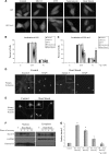
Figure 1.
Figure 1.
Heat shock increases nuclear localization of ataxin-3. (A) HeLa cells were transfected with GFP vector or GFP-Atx3 and 24 h later exposed to either 10 μm MG132 for 8 h, 1 mm DTT for 4 h, 10 μg/ml tunicamycin for 8 h, heat-shocked at 42°C for 1 h or maintained as untreated controls. Following treatment, GFP proteins were visualized in live cells. MG132, tunicamycin or DTT treatment did not alter localization of GFP-Atx3 or GFP. Heat shock did not affect localization of GFP; however, it increased nuclear localization of GFP-Atx3. Immunofluorescence data were quantified by counting GFP (B) or GFP-Atx3 (C) transfected cells with fluorescence primarily localized to the cytoplasm (C), nucleus (N) or evenly distributed (C/N). Data represent the mean ± SD of the fraction of total cells counted in each group with a particular localization pattern in three independent experiments with treated cells compared with controls (*P ≤ 0.01). Primary rat astrocytes (D) or primary rat cortical neurons (E) were either heat-shocked at 42°C for 30 min or left at 37°C. Following heat shock, cells were fixed and immunostained with Atx3 polyclonal antibody. In control cells, endogenous Atx3 localized throughout the cell, but after heat shock endogenous Atx3 redistributed to a primarily nuclear localization. Astrocytes were stained with DAPI to confirm the location of the nucleus. Cells indicated by white arrows are shown in insets. (F) Mouse fibroblasts were maintained at 37°C (C) or heat-shocked at 42°C for 3 h and collected over the following 24 h for cellular fractionation. Endogenous Atx3 localized to the nucleus following a 3 h heat shock and over the next 24 h returned to the cytoplasm. Distribution of GAPDH and histone H3 indicates purity of cytoplasmic and nuclear fractions. (G) Heat shock-induced nuclear accumulation of endogenous Atx3 was quantitated using densitometry. Data represent the mean ± SD of three independent experiments with data normalized to nuclear Atx3 levels in control cells. Nuclear levels of endogenous Atx3 increased 6.5-fold immediately following heat shock (P ≤ 0.01) and remained elevated following 2 h of recovery (P ≤ 0.01).
Figure 2.
Figure 2.
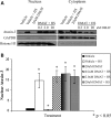
CK2 phosphorylation of ataxin-3 is not required for heat shock-induced nuclear localization. (A) HEK293T cells were treated with the CK2 inhibitor DMAT (0.2, 2.0 or 20 μm) or vehicle for 3 h and then heat-shocked for 1 h at 42°C and fractionated. Western blots indicated that following DMAT treatment, endogenous Atx3 still accumulated in the nucleus in response to heat shock similar to vehicle-treated cells. GAPDH and histone H3 were fractionation controls. (B) Heat shock-induced nuclear accumulation of endogenous Atx3 following vehicle or DMAT treatment was quantitated using densitometry. Data represent the mean ± SD of four independent experiments with all data normalized to vehicle control (*P < 0.05). No difference was detected between the DMAT + HS-treated groups and vehicle + HS group. DMAT treatment decreased the low level of nuclear Atx3 in cells maintained at 37°C by ∼60% (P < 0.05).
Figure 3.
Figure 3.
Effect of UIM and DUB mutations and polyglutamine expansion on heat shock-induced nuclear accumulation of ataxin-3. (A) HeLa cells transfected with myc-Atx3(Q29)C14A (DUB mutant) or myc-Atx3(Q29)S236/256D (UIM mutant) were either kept at 37°C or heat-shocked at 42°C for 1 h and immunostained with an myc antibody. (B) HEK293T cells transfected with myc-Atx3(Q29)C14A, or myc-Atx3(Q29)S236/256D, were either kept at 37°C or heat-shocked at 42°C for 1 h and nuclear and cytoplasmic fractions prepared. GAPDH and histone H3 were fractionation controls. (C) Heat shock-induced nuclear accumulation of Atx3(Q29)C14A and myc-Atx3(Q29)S236/256D were quantitated using densitometry. Data represent the mean ± SD of three independent experiments with each heat-shock group normalized to their 37°C control (*P < 0.05; **P < 0.01). Western blots and immunofluorescence data indicated that neither DUB activity nor functional UIMs were required for heat shock-induced nuclear localization of Atx3. (D) Pathogenic myc-Atx3(Q72) and WT myc-Atx3(Q29) were transfected into HeLa cells and either heat-shocked at 42°C for 1 h or kept at 37°C and immunostained with an myc antibody. (E) HEK293T cells transfected with myc-Atx3(Q29) or myc-Atx3(Q72) were heat-shocked at 42°C for 1 h or kept at 37°C and nuclear and cytoplasmic fractions prepared. In response to heat shock both wild-type and pathogenic Atx3 increased similarly in the nucleus. (F) Heat shock-induced nuclear accumulation of myc-Atx3(Q29) and myc-Atx3(Q72) were quantitated using densitometry. Data represent the mean ± SD of three independent experiments with data normalized to their 37°C control and displayed as fold increase in response to heat shock.
Figure 4.
Figure 4.
Effect of NLS mutation on heat shock-induced nuclear accumulation of ataxin-3. (A) HEK293T cells transfected with myc-Atx3 or the NLS mutant myc-Atx3(NLS*) were heat-shocked at 42°C for 1 h or kept at 37°C and nuclear and cytoplasmic fractions prepared. In response to heat shock Atx3 and Atx3(NLS*) increased similarly in the nucleus. (B) Heat shock-induced nuclear accumulation of myc-Atx3 and myc-Atx3(NLS*) were quantitated using densitometry. Data represent the mean ± SD of three independent experiments with data normalized to Atx3 control (*P < 0.05).
Figure 5.
Figure 5.
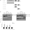
Mapping regions of ataxin-3 important for heat shock-induced nuclear localization. (A) Schematic of constructs used to map the regions of Atx3 involved in nuclear localization: full-length Atx3(Q29) (WT); Atx3(1–191) (N-terminal Josephin domain containing DUB activity); Atx3(191–362) (2 UIMs + PolyQ domain + C-terminus); Atx3(191–291) (2 UIMs) and Atx3(291–362) (PolyQ domain + C-terminus). (B) HEK293T cells were transfected with various myc-tagged Atx3 truncations to determine domains of Atx3 important for heat shock-induced nuclear localization. Both the N-terminal Josephin domain, Atx3(1–191), and the C-terminal half of the protein, Atx3(191–362), accumulated in the nucleus in response to heat shock, indicating that there were at least two sequences within Atx3 sufficient for nuclear localization. Atx3(291–362) also accumulated in the nucleus after heat shock, but Atx3(191–291) did not, suggesting that the second sequence important for regulating Atx3 heat shock-induced nuclear localization was located within amino acids 291–362. Atx3 truncations containing the polyQ domain typically migrate in SDS PAGE gels at multiple molecular weights that possibly represent multimers (these are identified with arrows and the number of potential ‘monomeric units’). (C) Heat shock-induced nuclear accumulation of each Atx3 truncation was quantitated using densitometry. Data represent the mean ± SD of three independent experiments with each heat-shock group normalized to their 37°C control (*P < 0.05).
Figure 6.
Figure 6.
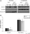
Ataxin-3 nuclear localization is regulated by serine-111 and independent of Hsf1. (A) Hsf1 WT and KO fibroblasts were either kept at 37°C or heat-shocked for 1 h at 42°C and cellular fractions prepared. Atx3 primarily localized to the nucleus after heat shock in both the Hsf1 WT and KO cells indicating that Hsf1 was not necessary for heat shock-induced nuclear localization of Atx3. GAPDH and histone H3 blots were controls for cell fractions. (B) Table showing the preferred sequence for Plk1 phosphorylation (52,53), several known substrates of Plk1 (Cyclin B1, Cdc25, Myt1, BRCA2 and Hsf1) and a potential phosphorylation site in Atx3 at serine-111. The sequence surrounding the Plk1 phosphorylation sites in known Plk1 substrates loosely resemble a predicted ‘preferred’ sequence. (C) HEK293T cells transfected with either Atx3, Atx3 S111A, Atx3(1–191) or Atx3(1–191) S111A were heat-shocked at 42°C for 1 h or kept at 37°C and then used for cellular fractionation. (D) Heat shock-induced nuclear accumulation of Atx3, Atx3 S111A, Atx3(1–191) and Atx3(1–191) S111A were quantitated using densitometry. Data represent the mean ± SD of three independent experiments comparing Atx3 with Atx3 S111A and Atx3(1–191) with Atx3(1–191) S111A (*P ≤ 0.05). Atx3 S111A and Atx3(1–191) S111A data were normalized to Atx3 and Atx3(1–191) data, respectively. Atx3 S111A showed a modest increase in the nucleus in response to heat shock but did not accumulate in the nucleus as much as wild-type Atx3. Nuclear localization of Atx3(1–191) increased following heat shock but Atx3(1–191) S111A did not accumulate in the nucleus in response to heat shock. (E) HEK293T cells transfected with either Atx3, Atx3 S111D, Atx3(1–191) or Atx3(1–191) S111D were heat-shocked at 42°C for 1 h or kept at 37°C and then used for cellular fractionation. Nuclear Atx3 S111D and Atx3(1–191) S111D increased in the nucleus in response to heat shock similar to Atx3 and Atx3(1–191). (F) Heat shock-induced nuclear accumulation of Atx3, Atx3 S111D, Atx3(1–191) and Atx3(1–191) S111D were quantitated using densitometry. Data represent the mean ± SD of three independent experiments. Atx3 S111D and Atx3(1–191) S111D data were normalized to Atx3 and Atx3(1–191) data, respectively. Atx3 S111D and Atx3(1–191) S111D increased in the nucleus similar to Atx3 and Atx3(1–191).
Figure 7.
Figure 7.
Ataxin-3 knockout fibroblasts are more sensitive to toxic heat shock. Atx3 WT and KO primary mouse fibroblasts were either (A) heat-shocked or treated with (B) MG132 or (C) DTT. Cell viability at each time point is expressed relative to the number of viable cells in parallel sets of control cultures. Data represent mean ± SD of three independent experiments; statistical comparisons are for WT versus KO cells at the same time point (*P < 0.05; **P < 0.01). (A) Atx3 WT and KO primary fibroblasts were heat-shocked at 45°C for 1 or 2 h or kept at 37°C as a control. Cell viability was measured immediately following heat shock and again 24 h later and represented as a percentage of control cells. Atx3 WT and KO primary mouse fibroblasts were treated with (B) 10μm MG132 for 8 or 12 h or (C) 2 mm DTT for 6 or 12 h and the number of viable cells were measured and represented as a percentage of control cells. Atx3 WT and KO fibroblasts responded similarly to MG132 and DTT treatment. (D) Atx3 WT and KO mouse fibroblasts were either kept at 37°C or heat-shocked at 42°C for 3 h and collected over the following 24 h for preparation of nuclear and cytoplasmic fractions. As expected no Atx3 was detected in Atx3 KO cells. In Atx3 WT cells endogenous Atx3 accumulated in the nucleus following heat shock and then returned to the cytoplasm between 4 and 24 h following heat shock. Both WT and KO fibroblasts initiated early events in the heat shock response resulting in Hsf1 translocating to the nucleus following heat shock as well as Hsf1 returning to the cytoplasm following induction of hsp70 expression.
Figure 8.
Figure 8.
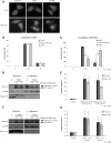
Wild-type and pathogenic ataxin-3 accumulate in the nucleus following oxidative stress. (A) HeLa cells were transfected with either GFP or GFP-Atx3 and exposed to 1 mm H2O2 for 5 h or 10 mm 3-NPA for 12 h. Following treatment GFP-Atx3 accumulated in the nucleus. Immunofluorescence data were quantitated by counting GFP (B) or GFP-Atx3 (C) transfected cells with fluorescence primarily localized to the cytoplasm (C), nucleus (N), or evenly distributed (C/N). Data represent the mean ± SD of the fraction of total cells counted in each group with a particular localization pattern in three independent experiments; treated cells were compared with control cells (*P < 0.01). (D) SCA3 patient fibroblasts expressing both WT and pathogenic Atx3 were exposed to 2.5 mm H2O2 for 4 h and cellular fractionations were performed following 0 or 4 h of recovery. Endogenous wild-type and pathogenic Atx3 accumulated in the nucleus of SCA3 patient fibroblasts following treatment with H2O2. (E) H2O2-induced nuclear accumulation of endogenous wild-type and pathogenic Atx3 were quantitated using densitometry. Data represent the mean ± SD of three independent experiments; for statistics H2O2- treated cells were compared with control cells (*P < 0.01). (F) SCA3 patient fibroblasts were exposed to 10 mm 3-NPA for 24 h and subcellular fractionations prepared following 0 or 4 h of recovery. Endogenous wild-type and pathogenic Atx3 accumulated in the nucleus of SCA3 patient fibroblasts following treatment with 3-NPA. (G) 3-NPA-induced nuclear accumulation of endogenous wild-type and pathogenic Atx3 was quantitated using densitometry. Data represent the mean ± SD of three independent experiments; 3-NPA-treated cells were compared with control cells (*P < 0.05).

Similar articles

Cited by

References

    1. Orr H.T., Zoghbi H.Y. Trinucleotide repeat disorders. Annu. Rev. Neurosci. 2007;30:575–621. - PubMed
    1. Paulson H.L., Perez M.K., Trottier Y., Trojanowski J.Q., Subramony S.H., Das S.S., Vig P., Mandel J.L., Fischbeck K.H., Pittman R.N. Intranuclear inclusions of expanded polyglutamine protein in spinocerebellar ataxia type 3. Neuron. 1997;19:333–344. - PubMed
    1. Davies S.W., Turmaine M., Cozens B.A., DiFiglia M., Sharp A.H., Ross C.A., Scherzinger E., Wanker E.E., Mangiarini L., Bates G.P. Formation of neuronal intranuclear inclusions underlies the neurological dysfunction in mice transgenic for the HD mutation. Cell. 1997;90:537–548. - PubMed
    1. Cummings C.J., Mancini M.A., Antalffy B., DeFranco D.B., Orr H.T., Zoghbi H.Y. Chaperone suppression of aggregation and altered subcellular proteasome localization imply protein misfolding in SCA1. Nat. Genet. 1998;19:148–154. - PubMed
    1. Perez M.K., Paulson H.L., Pendse S.J., Saionz S.J., Bonini N.M., Pittman R.N. Recruitment and the role of nuclear localization in polyglutamine-mediated aggregation. J. Cell Biol. 1998;143:1457–1470. - PMC - PubMed

Publication types

MeSH terms