

Lung adenocarcinoma with EGFR amplification has distinct clinicopathologic and molecular features in never-smokers
- PMID: 19826035
- PMCID: PMC2783286
- DOI: 10.1158/0008-5472.CAN-09-2477
Lung adenocarcinoma with EGFR amplification has distinct clinicopathologic and molecular features in never-smokers
Abstract
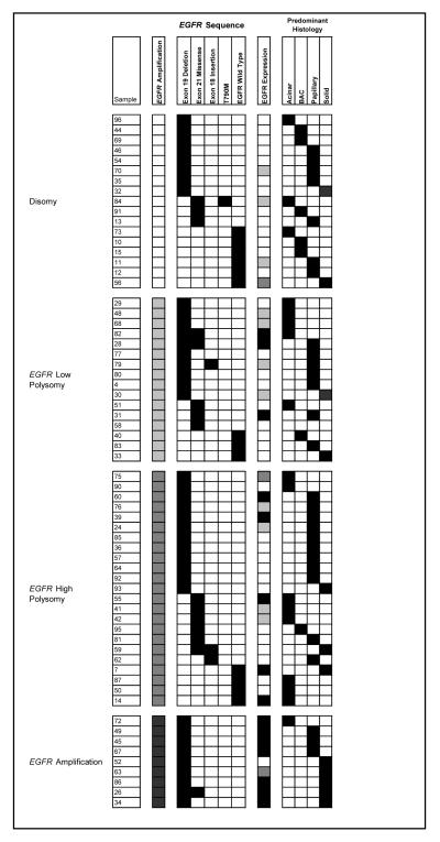
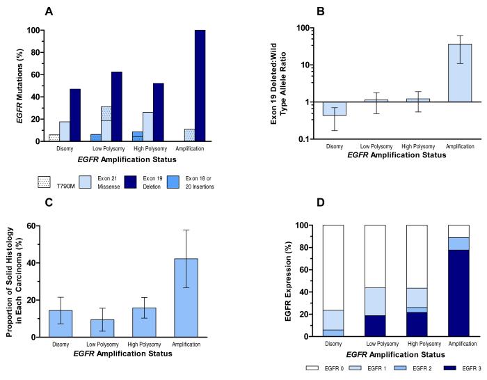
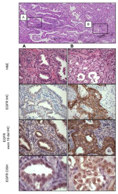
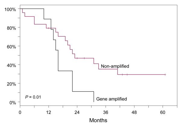
In a subset of lung adenocarcinomas, the epidermal growth factor receptor (EGFR) is activated by kinase domain mutations and/or gene amplification, but the interaction between the two types of abnormalities is complex and unclear. For this study, we selected 99 consecutive never-smoking women of East Asian origin with lung adenocarcinomas that were characterized by histologic subtype. We analyzed EGFR mutations by PCR-capillary sequencing, EGFR copy number abnormalities by fluorescence and chromogenic in situ hybridization and quantitative PCR, and EGFR expression by immunohistochemistry with both specific antibodies against exon 19 deletion-mutated EGFR and total EGFR. We compared molecular and clinicopathologic features with disease-free survival. Lung adenocarcinomas with EGFR amplification had significantly more EGFR exon 19 deletion mutations than adenocarcinomas with disomy, and low and high polysomy (100% versus 54%, P = 0.009). EGFR amplification occurred invariably on the mutated and not the wild-type allele (median mutated/wild-type ratios 14.0 versus 0.33, P = 0.003), was associated with solid histology (P = 0.008), and advanced clinical stage (P = 0.009). EGFR amplification was focally distributed in lung cancer specimens, mostly in regions with solid histology. Patients with EGFR amplification had a significantly worse outcome in univariate analysis (median disease-free survival, 16 versus 31 months, P = 0.01) and when adjusted for stage (P = 0.027). Lung adenocarcinomas with EGFR amplification have a unique association with exon 19 deletion mutations and show distinct clinicopathologic features associated with a significantly worsened prognosis. In these cases, EGFR amplification is heterogeneously distributed, mostly in areas with a solid histology.
Figures
Similar articles
-
Do all lung adenocarcinomas follow a stepwise progression?Lung Cancer. 2011 Oct;74(1):7-11. doi: 10.1016/j.lungcan.2011.05.021. Epub 2011 Jun 25. Lung Cancer. 2011. PMID: 21705107 Free PMC article. Review.
-
[Relationship between EGFR and KRAS mutations and prognosis in Chinese patients with non-small cell lung cancer: a mutation analysis with real-time polymerase chain reaction using scorpion amplification refractory mutation system].Zhonghua Bing Li Xue Za Zhi. 2012 Oct;41(10):652-6. doi: 10.3760/cma.j.issn.0529-5807.2012.10.002. Zhonghua Bing Li Xue Za Zhi. 2012. PMID: 23302304 Chinese.
-
Co-existence of positive MET FISH status with EGFR mutations signifies poor prognosis in lung adenocarcinoma patients.Lung Cancer. 2012 Jan;75(1):89-94. doi: 10.1016/j.lungcan.2011.06.004. Epub 2011 Jul 5. Lung Cancer. 2012. PMID: 21733594
-
Clinicopathologic and prognostic significance of c-MYC copy number gain in lung adenocarcinomas.Br J Cancer. 2014 May 27;110(11):2688-99. doi: 10.1038/bjc.2014.218. Epub 2014 May 8. Br J Cancer. 2014. PMID: 24809777 Free PMC article.
-
Pulmonary adenocarcinoma: a renewed entity in 2011.Respirology. 2012 Jan;17(1):50-65. doi: 10.1111/j.1440-1843.2011.02095.x. Respirology. 2012. PMID: 22040022 Free PMC article. Review.
Cited by
-
Structure-function analysis of oncogenic EGFR Kinase Domain Duplication reveals insights into activation and a potential approach for therapeutic targeting.Nat Commun. 2021 Mar 2;12(1):1382. doi: 10.1038/s41467-021-21613-6. Nat Commun. 2021. PMID: 33654076 Free PMC article.
-
Identifying a 6-Gene Prognostic Signature for Lung Adenocarcinoma Based on Copy Number Variation and Gene Expression Data.Oxid Med Cell Longev. 2022 Sep 28;2022:6962163. doi: 10.1155/2022/6962163. eCollection 2022. Oxid Med Cell Longev. 2022. Retraction in: Oxid Med Cell Longev. 2023 Dec 29;2023:9890283. doi: 10.1155/2023/9890283. PMID: 36211815 Free PMC article. Retracted.
-
Utility of Genomic Analysis in Differentiating Synchronous and Metachronous Lung Adenocarcinomas from Primary Adenocarcinomas with Intrapulmonary Metastasis.Transl Oncol. 2017 Jun;10(3):442-449. doi: 10.1016/j.tranon.2017.02.009. Epub 2017 Apr 25. Transl Oncol. 2017. PMID: 28448960 Free PMC article.
-
Elucidating Genomic Characteristics of Lung Cancer Progression from In Situ to Invasive Adenocarcinoma.Sci Rep. 2016 Aug 22;6:31628. doi: 10.1038/srep31628. Sci Rep. 2016. PMID: 27545006 Free PMC article.
-
Do all lung adenocarcinomas follow a stepwise progression?Lung Cancer. 2011 Oct;74(1):7-11. doi: 10.1016/j.lungcan.2011.05.021. Epub 2011 Jun 25. Lung Cancer. 2011. PMID: 21705107 Free PMC article. Review.
References
-
- Sharma SV, Bell DW, Settleman J, Haber DA. Epidermal growth factor receptor mutations in lung cancer. Nat Rev Cancer. 2007;7:169–81. - PubMed
-
- Lynch TJ, Bell DW, Sordella R, et al. Activating mutations in the epidermal growth factor receptor underlying responsiveness of non-small-cell lung cancer to gefitinib. N Engl J Med. 2004;350:2129–39. - PubMed
-
- Paez JG, Janne PA, Lee JC, et al. EGFR mutations in lung cancer: correlation with clinical response to gefitinib therapy. Science. 2004;304:1497–500. - PubMed
-
- Huang SF, Liu HP, Li LH, et al. High frequency of epidermal growth factor receptor mutations with complex patterns in non-small cell lung cancers related to gefitinib responsiveness in Taiwan. Clin Cancer Res. 2004;10:8195–203. - PubMed
-
- Shigematsu H, Lin L, Takahashi T, et al. Clinical and biological features associated with epidermal growth factor receptor gene mutations in lung cancers. J Natl Cancer Inst. 2005;97:339–46. - PubMed
Publication types
MeSH terms
Substances
Grants and funding
LinkOut - more resources
Full Text Sources
Other Literature Sources
Medical
Research Materials
Miscellaneous
