

The knockout of miR-143 and -145 alters smooth muscle cell maintenance and vascular homeostasis in mice: correlates with human disease
- PMID: 19816508
- PMCID: PMC3014107
- DOI: 10.1038/cdd.2009.153
The knockout of miR-143 and -145 alters smooth muscle cell maintenance and vascular homeostasis in mice: correlates with human disease
Abstract
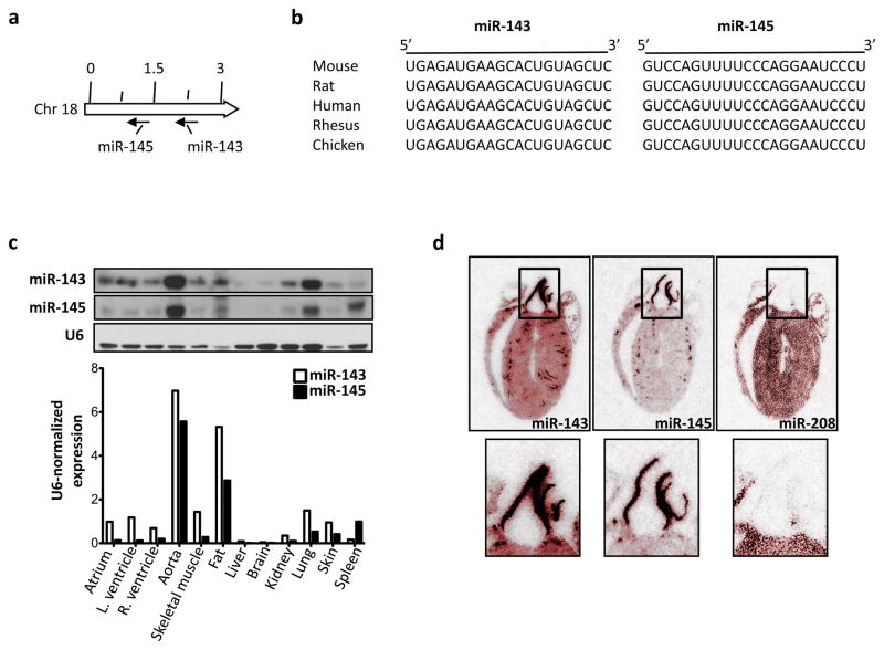
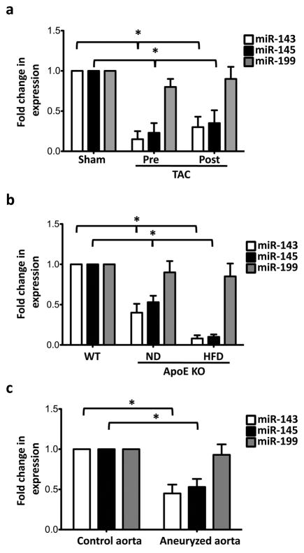
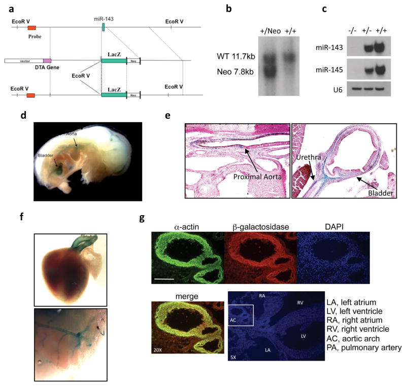
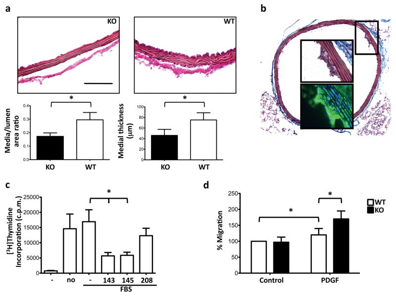
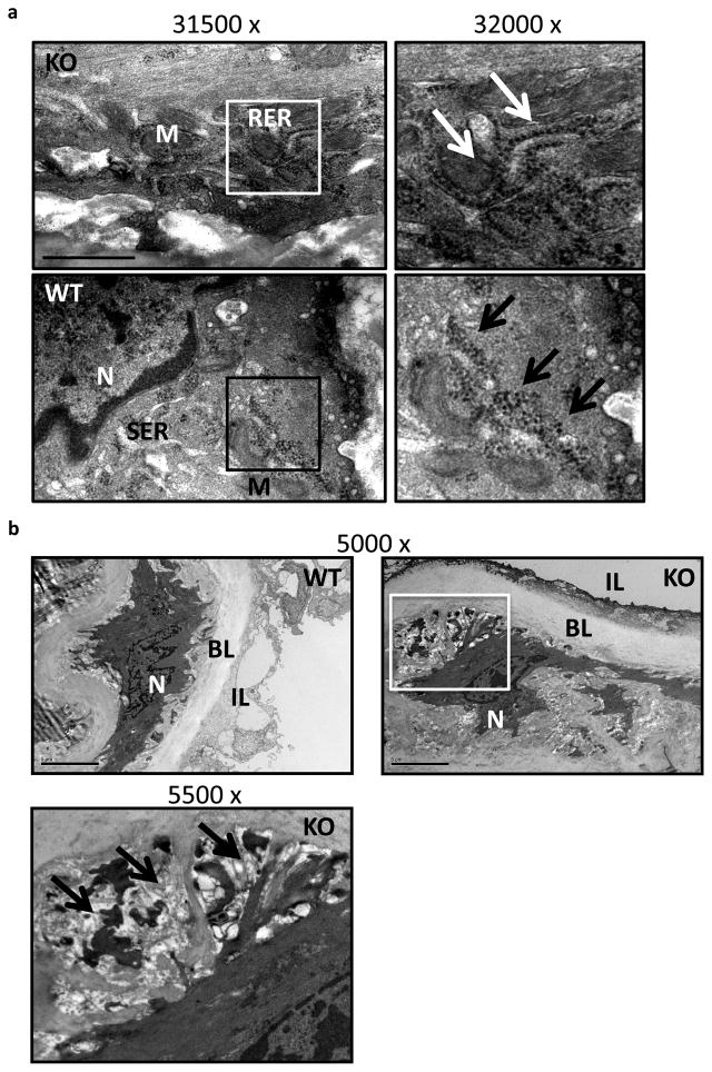
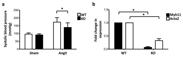
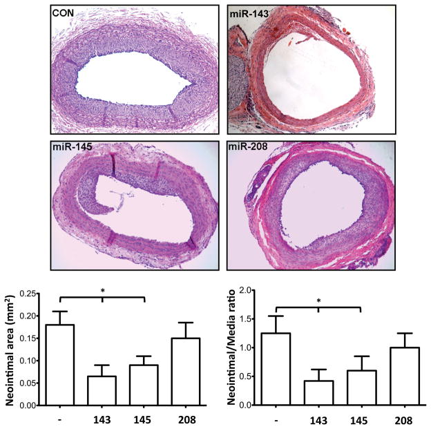
Mechanisms controlling vascular smooth muscle cell (VSMC) plasticity and renewal still remain to be elucidated completely. A class of small RNAs called microRNAs (miRs) regulate gene expression at the post-transcriptional level. Here, we show a critical role of the miR-143/145 cluster in SMC differentiation and vascular pathogenesis, also through the generation of a mouse model of miR-143 and -145 knockout (KO). We determined that the expression of miR-143 and -145 is decreased in acute and chronic vascular stress (transverse aortic constriction and in aortas of the ApoE KO mouse). In human aortic aneurysms, the expression of miR-143 and -145 was significantly decreased compared with control aortas. In addition, overexpression of miR-143 and -145 decreased neointimal formation in a rat model of acute vascular injury. An in-depth analysis of the miR-143/145 KO mouse model showed that this miR cluster is expressed mostly in the SMC compartment, both during development and postnatally, in vessels and SMC-containing organs. Loss of miR-143 and miR-145 expression induces structural modifications of the aorta, because of an incomplete differentiation of VSMCs. In conclusion, our results show that the miR-143/145 gene cluster has a critical role during SMC differentiation and strongly suggest its involvement in the reversion of the VSMC differentiation phenotype that occurs during vascular disease.
Figures
Similar articles
-
Oncological miR-182-3p, a Novel Smooth Muscle Cell Phenotype Modulator, Evidences From Model Rats and Patients.Arterioscler Thromb Vasc Biol. 2016 Jul;36(7):1386-97. doi: 10.1161/ATVBAHA.115.307412. Epub 2016 May 19. Arterioscler Thromb Vasc Biol. 2016. PMID: 27199451
-
Down-regulation of miR-23b induces phenotypic switching of vascular smooth muscle cells in vitro and in vivo.Cardiovasc Res. 2015 Sep 1;107(4):522-33. doi: 10.1093/cvr/cvv141. Epub 2015 May 20. Cardiovasc Res. 2015. PMID: 25994172
-
miR-128-3p Is a Novel Regulator of Vascular Smooth Muscle Cell Phenotypic Switch and Vascular Diseases.Circ Res. 2020 Jun 5;126(12):e120-e135. doi: 10.1161/CIRCRESAHA.120.316489. Epub 2020 Mar 27. Circ Res. 2020. PMID: 32216529
-
Smooth muscle miRNAs are critical for post-natal regulation of blood pressure and vascular function.PLoS One. 2011 Apr 22;6(4):e18869. doi: 10.1371/journal.pone.0018869. PLoS One. 2011. PMID: 21526127 Free PMC article.
-
Can microRNAs control vascular smooth muscle phenotypic modulation and the response to injury?Physiol Genomics. 2011 May 1;43(10):529-33. doi: 10.1152/physiolgenomics.00146.2010. Epub 2010 Sep 14. Physiol Genomics. 2011. PMID: 20841497 Free PMC article. Review.
Cited by
-
MicroRNAs in pulmonary arterial remodeling.Cell Mol Life Sci. 2013 Dec;70(23):4479-94. doi: 10.1007/s00018-013-1382-5. Epub 2013 Jun 6. Cell Mol Life Sci. 2013. PMID: 23739951 Free PMC article. Review.
-
The role of miR-143/miR-145 in the development, diagnosis, and treatment of diabetes.J Diabetes Metab Disord. 2023 Oct 11;23(1):39-47. doi: 10.1007/s40200-023-01317-y. eCollection 2024 Jun. J Diabetes Metab Disord. 2023. PMID: 38932869 Free PMC article. Review.
-
Regulation of microRNAs by Brahma-related gene 1 (Brg1) in smooth muscle cells.J Biol Chem. 2013 Mar 1;288(9):6397-408. doi: 10.1074/jbc.M112.409474. Epub 2013 Jan 20. J Biol Chem. 2013. PMID: 23339192 Free PMC article.
-
microRNA-based diagnostics and therapy in cardiovascular disease-Summing up the facts.Cardiovasc Diagn Ther. 2015 Feb;5(1):17-36. doi: 10.3978/j.issn.2223-3652.2014.12.03. Cardiovasc Diagn Ther. 2015. PMID: 25774345 Free PMC article. Review.
-
Induction of microRNA-1 by myocardin in smooth muscle cells inhibits cell proliferation.Arterioscler Thromb Vasc Biol. 2011 Feb;31(2):368-75. doi: 10.1161/ATVBAHA.110.218149. Epub 2010 Nov 4. Arterioscler Thromb Vasc Biol. 2011. PMID: 21051663 Free PMC article.
References
-
- Gimona M, Herzog M, Vandekerckhove J, Small JV. Smooth muscle specific expression of calponin. FEBS Lett. 1990 Nov 12;274:159–162. - PubMed
-
- Sobue K, Hayashi K, Nishida W. Expressional regulation of smooth muscle cell-specific genes in association with phenotypic modulation. Mol Cell Biochem. 1999 Jan;190:105–118. - PubMed
-
- Schwartz SM. Smooth muscle migration in atherosclerosis and restenosis. J Clin Invest. 1997 Dec 1;100:S87–89. - PubMed
-
- Eberhard A, Kahlert S, Goede V, Hemmerlein B, Plate KH, Augustin HG. Heterogeneity of angiogenesis and blood vessel maturation in human tumors: implications for antiangiogenic tumor therapies. Cancer Res. 2000 Mar 1;60:1388–1393. - PubMed
-
- Owens GK, Kumar MS, Wamhoff BR. Molecular regulation of vascular smooth muscle cell differentiation in development and disease. Physiol Rev. 2004 Jul;84:767–801. - PubMed
Publication types
MeSH terms
Substances
Grants and funding
LinkOut - more resources
Full Text Sources
Other Literature Sources
Molecular Biology Databases
Research Materials
Miscellaneous
