Temporomandibular joint formation requires two distinct hedgehog-dependent steps
- PMID: 19815519
- PMCID: PMC2775291
- DOI: 10.1073/pnas.0908836106
Temporomandibular joint formation requires two distinct hedgehog-dependent steps
Abstract
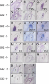
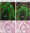
We conducted a genetic analysis of the developing temporo-mandibular or temporomandi-bular joint (TMJ), a highly specialized synovial joint that permits movement and function of the mammalian jaw. First, we used laser capture microdissection to perform a genome-wide expression analysis of each of its developing components. The expression patterns of genes identified in this screen were examined in the TMJ and compared with those of other synovial joints, including the shoulder and the hip joints. Striking differences were noted, indicating that the TMJ forms via a distinct molecular program. Several components of the hedgehog (Hh) signaling pathway are among the genes identified in the screen, including Gli2, which is expressed specifically in the condyle and in the disk of the developing TMJ. We found that mice deficient in Gli2 display aberrant TMJ development such that the condyle loses its growth-plate-like cellular organization and no disk is formed. In addition, we used a conditional strategy to remove Smo, a positive effector of the Hh signaling pathway, from chondrocyte progenitors. This cell autonomous loss of Hh signaling allows for disk formation, but the resulting structure fails to separate from the condyle. Thus, these experiments establish that Hh signaling acts at two distinct steps in disk morphogenesis, condyle initiation, and disk-condyle separation and provide a molecular framework for future studies of the TMJ.
Conflict of interest statement
The authors declare no conflict of interest.
Figures


Similar articles
-
Temporomandibular joint formation and condyle growth require Indian hedgehog signaling.Dev Dyn. 2007 Feb;236(2):426-34. doi: 10.1002/dvdy.21036. Dev Dyn. 2007. PMID: 17191253
-
Genetic Influences on Temporomandibular Joint Development and Growth.Curr Top Dev Biol. 2015;115:85-109. doi: 10.1016/bs.ctdb.2015.07.008. Epub 2015 Oct 1. Curr Top Dev Biol. 2015. PMID: 26589922 Review.
-
Trps1 is necessary for normal temporomandibular joint development.Cell Tissue Res. 2012 Apr;348(1):131-40. doi: 10.1007/s00441-012-1372-1. Epub 2012 Mar 17. Cell Tissue Res. 2012. PMID: 22427063
-
Spry1 and spry2 are essential for development of the temporomandibular joint.J Dent Res. 2012 Apr;91(4):387-93. doi: 10.1177/0022034512438401. Epub 2012 Feb 10. J Dent Res. 2012. PMID: 22328578 Free PMC article.
-
Osteophyte formation and matrix mineralization in a TMJ osteoarthritis mouse model are associated with ectopic hedgehog signaling.Matrix Biol. 2016 May-Jul;52-54:339-354. doi: 10.1016/j.matbio.2016.03.001. Epub 2016 Mar 3. Matrix Biol. 2016. PMID: 26945615 Free PMC article. Review.
Cited by
-
Muenke syndrome mutation, FgfR3P²⁴⁴R, causes TMJ defects.J Dent Res. 2012 Jul;91(7):683-9. doi: 10.1177/0022034512449170. Epub 2012 May 23. J Dent Res. 2012. PMID: 22622662 Free PMC article.
-
Clinical findings in patients with GLI2 mutations--phenotypic variability.Clin Genet. 2012 Jan;81(1):70-5. doi: 10.1111/j.1399-0004.2010.01606.x. Epub 2011 Jan 19. Clin Genet. 2012. PMID: 21204792 Free PMC article.
-
ScxLin cells directly form a subset of chondrocytes in temporomandibular joint that are sharply increased in Dmp1-null mice.Bone. 2021 Jan;142:115687. doi: 10.1016/j.bone.2020.115687. Epub 2020 Oct 12. Bone. 2021. PMID: 33059101 Free PMC article.
-
Evidence of vasculature and chondrocyte to osteoblast transdifferentiation in craniofacial synovial joints: Implications for osteoarthritis diagnosis and therapy.FASEB J. 2020 Mar;34(3):4445-4461. doi: 10.1096/fj.201902287R. Epub 2020 Feb 6. FASEB J. 2020. PMID: 32030828 Free PMC article.
-
Roles of Ihh signaling in chondroprogenitor function in postnatal condylar cartilage.Matrix Biol. 2018 Apr;67:15-31. doi: 10.1016/j.matbio.2018.02.011. Epub 2018 Feb 12. Matrix Biol. 2018. PMID: 29447948 Free PMC article.
References
-
- Avery JK. Oral Development and Histology. 3rd Ed. New York: Thieme; 2001. p. 435.
-
- Hinchliffe JR, Johnson DR. The Development of the Vertebrate Limb. Oxford: Clarendon; 1980.
-
- Shubin NH, Alberch P. A morphogenetic approach to the origin and basic organization of the tetrapod limb. Evol Biol. 1986;20:319–387.
-
- Oster GF, Shubin N, Murray JD, Alberch P. Evolution and morphogenic rules: The shape of the vertebrate limb in ontogeny and phylogeny. Evolution. 1988;42:862–884. - PubMed
-
- Craig FM, Bentley G, Archer CW. The spatial and temporal pattern of collagens I and II and keratan sulphate in the developing chick metatarsophalangeal joint. Development. 1987;99:383–391. - PubMed
Publication types
MeSH terms
Substances
Grants and funding
LinkOut - more resources
Full Text Sources
Molecular Biology Databases
Miscellaneous

