Review
. 2009:335:1-32.
doi: 10.1007/978-3-642-00302-8_1.

An overview of the molecular mechanism of autophagy

Affiliations
Review

An overview of the molecular mechanism of autophagy

Zhifen Yang et al. Curr Top Microbiol Immunol. 2009.

Abstract

Autophagy is a highly conserved cellular degradation process in which portions of cytosol and organelles are sequestered into a double-membrane vesicle, an autophagosome, and delivered into a degradative organelle, the vacuole/lysosome, for breakdown and eventual recycling of the resulting macromolecules. This process relieves the cell from various stress conditions. Autophagy plays a critical role during cellular development and differentiation, functions in tumor suppression, and may be linked to life span extension. Autophagy also has diverse roles in innate and adaptive immunity, such as resistance to pathogen invasion. Substantial progress has been made in the identification of many autophagy-related (ATG) genes that are essential to drive this cellular process, including both selective and nonselective types of autophagy. Identification of the ATG genes in yeast, and the finding of orthologs in other organisms, reveals the conservation of the autophagic machinery in all eukaryotes. Here, we summarize our current knowledge about the machinery and molecular mechanism of autophagy.


Figures

Fig. 1
Fig. 1
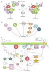
Schematic overview of autophagy and the Cvt pathway in yeast. Both pathways involve the engulfment of cargos within distinct double-membrane vesicles, which are thought to originate from the phagophore assembly site (PAS). The Cvt pathway is biosynthetic and is used for the delivery of two resident vacuolar hydrolases, aminopeptidase I (Ape1), and α-mannosidase (Ams1), and it occurs under vegetative conditions. The Cvt vesicle is approximately 140–160 nm in diameter and appears to closely enwrap the specific cargo, the Cvt complex (consisting of prApe1 and the Atg19 receptor), and exclude bulk cytoplasm. In contrast, autophagy is degradative and is induced by inactivation of Tor kinase upon nutrient starvation. The autophagosome, which is 300–900 nm in diameter, sequesters cytoplasm, including organelles, and can also specifically sequester the Cvt complex. Once completed, the double-membrane vesicles dock and fuse with the vacuole, and release the inner single-membrane vesicles (autophagic or Cvt body) into the lumen. Subsequently, these vesicles are broken down, allowing the maturation of prApe1 and the degradation of cytoplasm, with recycling of the resulting macromolecules through vacuolar permeases. This figure is modified from Fig. 1 of Yorimitsu and Klionsky (2005b)
Fig. 2
Fig. 2
Schematic representation of autophagy and autophagy-related pathways. These dynamic pathways can be broken down into a series of steps including induction, cargo recognition and packaging, vesicle nucleation, vesicle expansion and completion, Atg protein cycling, vesicle fusion with the vacuole/lysosome, vesicle breakdown and recycling of the resulting macromolecules. The Atg proteins can be classified into several different groups according to their functions at the different steps of the pathway. The Atg1 complex may act at multiple steps of the pathway, including induction and Atg protein cycling. During the vesicle formation process, several Atg proteins are involved in cycling between the peripheral sites and the PAS. PAS, phagophore assembly site; thought to be the organizing site for phagophore formation. This figure is modified from Fig. 2 of Huang and Klionsky (2007)
Fig. 3a–c
Fig. 3a–c
Regulation of autophagy induction in yeast and mammalian cells. a Regulatory complex for autophagy induction in yeast. In yeast, autophagy is mainly a starvation response, and Tor kinase complex 1 (TORC1) regulates the induction of autophagy upon sensing the nutrient conditions. Atg1 kinase, which is essential for both autophagy and the Cvt pathway, forms a putative complex with several Atg proteins that are primarily required for autophagy (in green) or the Cvt pathway (in purple). Under nutrient-rich conditions, TORC1 is active and Atg13 is highly phosphorylated, and this hyperphosphorylated Atg13 has a lower affinity for Atg1 and Atg17. Upon inactivation of TORC1 by nutrient starvation, Atg13 is rapidly and partially dephosphorylated, leading to a higher affinity for Atg1 and Atg17. The formation of the Atg1–Atg13–Atg17 ternary complex allows the activation of Atg1 kinase activity, which may regulate the switch between autophagy and the Cvt pathway in response to environmental changes. The function of additional components of the putative complex depicted here, including Atg20, Atg24, Atg29, Atg31 and Vac8, are not known. Atg11 may function in part as a scaffold protein. This figure is modified from Fig. 2 of Yorimitsu and Klionsky (2005b) b Multiple nutrient-sensing kinase signaling pathways converge on autophagy in yeast. TORC1 plays a major role in the regulation of autophagy. Ras is active under nutrient-rich conditions and allows the activation of PKA, which inhibits autophagy. The PKA and Sch9 signaling pathways cooperatively regulate the induction of autophagy in parallel with Tor, although Sch9 is also a direct substrate of TORC1. The eIF2α kinase signaling pathway positively regulates autophagy, and Gcn2 might be another target of TORC1. Snf1 and Pho85 are additional positive and negative regulatory components, respectively, of autophagy in yeast. c Regulation of autophagy in mammalian cells. mTor activation depends on several inputs, including nutrients (amino acids), energy (ATP) and growth factor (insulin/IGF). In response to insulin receptor stimulation, a class I phosphoinositide 3-kinase (PtdIns3K) is activated and generates PtdIns(3,4)P2 and PtdIns(3,4,5)P3 at the plasma membrane, and the latter two activate 3-phosphoinositide-dependent protein kinase 1 (PDK1) and protein kinase B (PKB)/Akt. PKB phosphorylates and inhibits the GTPase-activating protein complex TSC1–TSC2, leading to the stabilization of Rheb-GTP, which stimulates mTor, causing inhibition of autophagy. PTEN, a 3′-phosphoinositide phosphatase, antagonizes PKB and has a stimulatory effect on autophagy. Both mTor and PDK1 stimulate p70S6 kinase (p70S6K). In one model, under nutrient-rich conditions, activation of S6K directly stimulates autophagy, or it is stimulated indirectly through inhibition of PtdIns3K, allowing a basal level of autophagy for homeostatic purposes. Under starvation conditions, inhibition of mTor prevents further activation of S6K, which limits and prevents excessive autophagy. Both ATP and amino acid deprivation result in mTor inactivation independent of the insulin signaling pathway. Amino acids activate mTor via inhibition of the TSC1–TSC2 complex or are sensed by mTor directly. Energy stress causes activation of the LKB1–AMPK pathway, which inhibits mTor by activating TSC1–TSC2. AMPK phosphorylates and stabilizes p27, a cyclin-kinase inhibitor, leading to activation of autophagy. An antiapoptotic protein, Bcl-2, associates with Beclin 1, the mammalian homolog of Atg6, and inhibits a class III PtdIns3K complex, whereas the latter serves a stimulatory role in autophagy. Also shown is the notion that Atg1 overexpression negatively feeds back on Tor activity in Drosophila
Fig. 4
Fig. 4
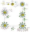
Temporal order of action of cargo recognition, packaging and sequestration in the Cvt pathway. A selective type of autophagy, the Cvt pathway, specifically transports the vacuolar hydrolases precursor Ape1 and Ams1 into the vacuole. Precursor Ape1 is synthesized in the cytosol, assembled into a dodecamer, and then further packaged into a larger oligomeric structure, called the Ape1 complex. Atg19 binds to the propeptide of prApe1 to form the Cvt complex in the cytosol; Ams1 is also incorporated into this complex via binding to Atg19. Atg11 subsequently associates with Atg19, acting as an adapter to bring the Cvt complex to the phagophore assembly site or PAS, a potential site for Cvt vesicle formation. The PAS may organize the formation of the sequestering vesicle, or it may literally become the sequestering vesicle as shown. Atg11 assembles with the Cvt complex before targeting to the PAS, and it forms a homodimer or homo-oligomer at the PAS, although it is not clear whether this self-interaction occurs before or after the arrival at this site. Several Atg components, including Atg8, are recruited to the PAS independent of Atg11. Atg8 is conjugated into Atg8—PE for subsequent vesicle formation. Atg8—PE interacts with Atg19, and allows the correct incorporation of the Cvt complex into the forming vesicle. Atg19 is delivered into the vacuole together with the cargo proteins and degraded there. The scaffold protein Atg11, however, dissociates from the Cvt complex before vesicle completion, although the exact timing and mechanism of its release remain to be resolved. This figure is modified from Fig. 3 of Yorimitsu and Klionsky (2005b)
Fig. 5
Fig. 5
Two phosphatidylinositol 3-kinase (PtdIns3K) complexes in yeast. Each complex contains three common components, Vps15, Vps34, and Vps30/Atg6. Vps34 is the PtdIns3K enzyme, and Vps15 is thought to be a regulatory component; the function of Vps30/Atg6 is not known. In addition, each complex contains another specific component, Atg14 (complex I) or Vps38 (complex II), which is thought to act as a connector between Vps30 and Vps15–Vps34. Complex I functions in autophagy and the Cvt pathway, whereas complex II acts in the Vps pathway, including the CPY and MVB pathways. This figure is modified from Fig. 5a of Yorimitsu and Klionsky (2005b)
Fig. 6
Fig. 6
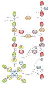
Two ubiquitin-like protein conjugation systems. The conjugation of Atg12 to Atg5 starts with activation by Atg7, which is homologous to the E1 ubiquitin-activating enzyme. Atg7 hydrolyzes ATP, resulting in the activation of Atg12 via the formation of a thioester bond between the C-terminal glycine of Atg12 and the active site cysteine of Atg7; subsequently, the activated Atg12 is transferred to the active site cysteine of Atg10, an E2-like enzyme, which catalyzes the conjugation of Atg12 to Atg5 through the formation of an isopeptide bond between the activated glycine of Atg12 and an internal lysine residue of Atg5. Atg12—Atg5 is finally assembled with Atg16. Atg16 forms a tetramer to allow the formation of an Atg12—Atg5—Atg16 multimeric structure. The conjugation of Atg8—PE starts with the cleavage of the C-terminal arginine of Atg8 by the protease Atg4. The exposed glycine of Atg8 is then bound to the active site cysteine of the same E1-like enzyme, Atg7. The activated Atg8 is then transferred to another E2-like enzyme, Atg3. Eventually, Atg3 catalyzes the conjugation of Atg8 to form Atg8—PE. The Atg12—Atg5 conjugate might function as an E3, ubiquitin ligase-like enzyme, to promote Atg8—PE conjugation. Both the Atg12—Atg5—Atg16 complex and Atg8—PE localize to the PAS to facilitate vesicle formation. The Atg8—PE that resides on the outer face of the sequestering vesicle is released from the membrane by a second Atg4-dependent cleavage. This figure is modified from Fig. 4 of Yorimitsu and Klionsky (2005b)
Fig. 7
Fig. 7
Cycling of Atg9. In yeast, Atg9 cycles between the PAS and non-PAS punctate structures (peripheral sites), some of which are found to localize adjacent to or at the surface of mitochondria. The efficient anterograde movement of Atg9 to the PAS requires Atg11, Atg23, Atg27, and the actin cytoskeletion. Atg9, Atg23, and Atg27 are in a heterotrimeric complex, and their movement to the PAS is interdependent. Atg11 acts as a potential adaptor between Atg9 and actin, and between Atg9 and the Arp2/3 complex, while the latter may provide the force to push the cargo (Atg9 and its associated membrane) away from the peripheral sites and toward the PAS. The retrograde transport of Atg9 from the PAS back to the peripheral sites depends on the Atg1–Atg13 kinase complex, Atg2, Atg18, and the PtdIns3K complex I. The Atg1–Atg13 complex promotes the association of Atg2 and the PtdIns(3)P binding protein Atg18 with Atg9, and the formation of this ternary complex initiates Atg9 retrieval for another round of membrane delivery
Fig. 8
Fig. 8
Schematic depiction of double-membrane vesicle formation. The PAS serves to facilitate the nucleation and/or expansion of the phagophore, the precursor of the autophagosome, through recruitment of Atg proteins. Atg9 and the PtdIns3K complex I are recruited relatively early to the PAS, and act in membrane nucleation. Atg9 cycles between peripheral sites and the PAS; potentially, Atg9 cycling delivers lipids to the expanding membrane. The Atg12—Atg5—Atg16 complex and the Atg8—PE conjugate, components of the vesicle-forming machinery, are subsequently recruited to the PAS, and mediate the expansion of the sequestering vesicle. The Atg12—Atg5—Atg16 complex may in part behave like a coat or may function as an E3 ubiquitin ligase-like enzyme, whereas Atg8—PE acts in the elongation of the vesicle as a structural component. Before or immediately after the autophagosome is completed, most of the Atg components, including the putative coat proteins, dissociate from the vesicle; the portion of Atg8—PE on the outer surface of the vesicle is normally cleaved off by the Atg4 protease. Finally, the sequestering vesicle can fuse with the vacuole
Fig. 9
Fig. 9
Vesicle docking and fusion with the vacuole. The SNARE proteins Vam3, Vam7, Vti1, and Ykt6 function in various membrane fusion events, including the process of autophagosome fusion with the vacuole. Also shown are the Mon1–Ccz1 complex and the class C Vps/HOPS complex, which function in concert at the Ypt7-dependent tethering/docking stage. This figure is modified from Fig. 7 of Klionsky (2005)

Similar articles

Cited by

References

    1. Abeliovich H, Zhang C, Dunn WA, Jr, Shokat KM, Klinosy DJ. Chemical genetic analysis of Apgl reveals a non-kinase role in the induction of autophagy. Mol Biol Cell. 2003;14:477–490. - PMC - PubMed
    1. Arico S, Petiot A, Bauvy C, Dubbelhuis PF, Meijer AJ, Codogno P, Ogier-Denis E. The tumor suppressor PTEN positively regulates macroautophagy by inhibiting the phosphatidylinositol 3-kinase/protein kinase B pathway. J Biol Chem. 2001;276:35243–35246. - PubMed
    1. Atlashkin V, Kreykenbohm V, Eskelinen EL, Wenzel D, Fayyazi A, Fischer von Mollard G. Deletion of the SNARE vti1b in mice results in the loss of a single SNARE partner, syntaxin 8. Mol Cell Biol. 2003;23:5198–5207. - PMC - PubMed
    1. Baba M, Osumi M, Scott SV, Klionsky DJ, Ohsumi Y. Two distinct pathways for targeting proteins from the cytoplasm to the vacuole/lysosome. J Cell Biol. 1997;139:1687–1695. - PMC - PubMed
    1. Bellu AR, Komori M, van der Klei IJ, Kiel JAKW, Veenhuis M. Peroxisome biogenesis and selective degradation converge at Pex14p. J Biol Chem. 2001;276:44570–44574. - PubMed

Publication types

LinkOut - more resources