. 2010 Apr;51(4):709-19.
doi: 10.1194/jlr.M001917. Epub 2009 Oct 2.

CGI-58/ABHD5 is a coenzyme A-dependent lysophosphatidic acid acyltransferase

Affiliations

CGI-58/ABHD5 is a coenzyme A-dependent lysophosphatidic acid acyltransferase

Gabriela Montero-Moran et al. J Lipid Res. 2010 Apr.

Abstract

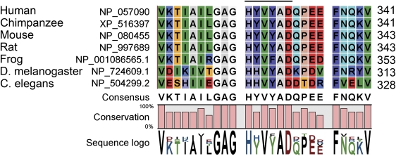
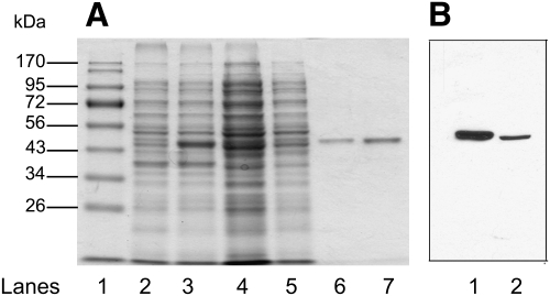
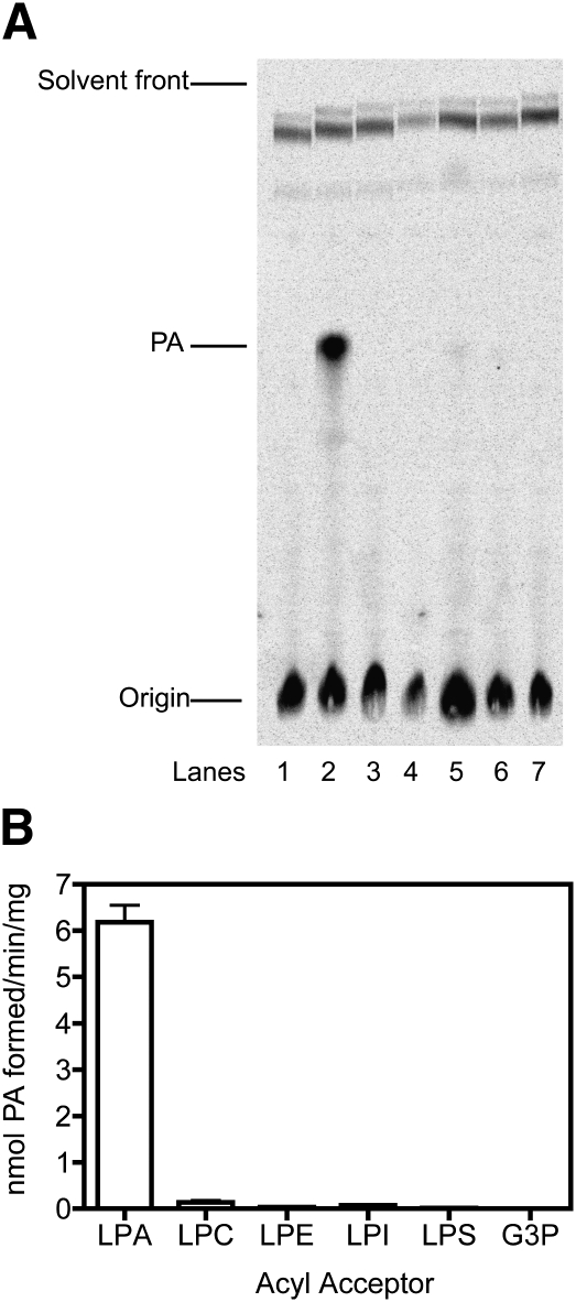
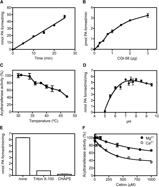
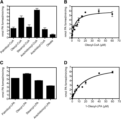
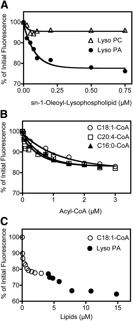
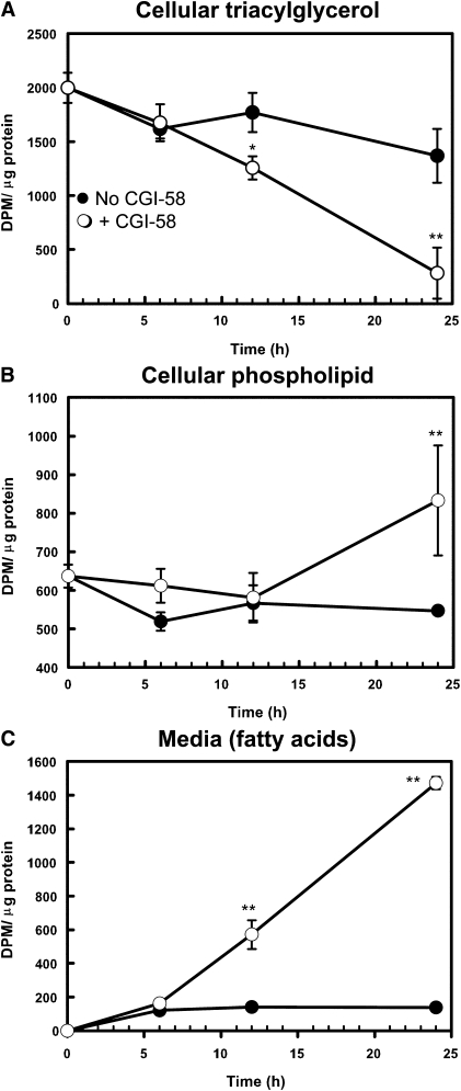
Mutations in human CGI-58/ABHD5 cause Chanarin-Dorfman syndrome (CDS), characterized by excessive storage of triacylglycerol in tissues. CGI-58 is an alpha/beta-hydrolase fold enzyme expressed in all vertebrates. The carboxyl terminus includes a highly conserved consensus sequence (HXXXXD) for acyltransferase activity. Mouse CGI-58 was expressed in Escherichia coli as a fusion protein with two amino terminal 6-histidine tags. Recombinant CGI-58 displayed acyl-CoA-dependent acyltransferase activity to lysophosphatidic acid, but not to other lysophospholipid or neutral glycerolipid acceptors. Production of phosphatidic acid increased with time and increasing concentrations of recombinant CGI-58 and was optimal between pH 7.0 and 8.5. The enzyme showed saturation kinetics with respect to 1-oleoyl-lysophosphatidic acid and oleoyl-CoA and preference for arachidonoyl-CoA and oleoyl-CoA. The enzyme showed slight preference for 1-oleoyl lysophosphatidic acid over 1-palmitoyl, 1-stearoyl, or 1-arachidonoyl lysophosphatidic acid. Recombinant CGI-58 showed intrinsic fluorescence for tryptophan that was quenched by the addition of 1-oleoyl-lysophosphatidic acid, oleoyl-CoA, arachidonoyl-CoA, and palmitoyl-CoA, but not by lysophosphatidyl choline. Expression of CGI-58 in fibroblasts from humans with CDS increased the incorporation of radiolabeled fatty acids released from the lipolysis of stored triacylglycerols into phospholipids. CGI-58 is a CoA-dependent lysophosphatidic acid acyltransferase that channels fatty acids released from the hydrolysis of stored triacylglycerols into phospholipids.


Figures

Fig. 1.
Fig. 1.
Sequence alignment of amino acid sequences of CGI-58 from various species showing the conserved acyltransferase motif. Amino acid sequences for a portion of the carboxyl terminus of CGI-58 are aligned for human (NP_057090), chimpanzee (XP_516397), mouse (NP_080455), rat (NP_997689), frog (Xenopus laevis, NP_001086565.1), Drosophila melanogaster (CG1882, isoform B, NP_724609.1), and Caenorhabditis elegans (hypothetical protein C37H5.2, NP_504299.2) using CLC Genomics Workbench 5.1. The sequences show the conservation of the HXXXXD consensus site for acyltransferase residues, indicated by the over-scoring line. Numbers following each sequence refer to the final residue.
Fig. 2.
Fig. 2.
Purification of recombinant CGI-58. Recombinant His12-CGI-58 was expressed in E. coli and purified over Ni2+-NTA-agarose. A: Coomassie Blue-stained SDS-PAGE gel showing molecular weight markers (lane 1), uninduced E. coli lysate (lane 2), isopropyl-β-D-1-thiogalactopyranoside-induced bacterial lysate (lane 3), supernatant following cell disruption by French press (lane 4), supernatant from the polyethyleneimine precipitation (lane 5), and eluant from Ni2+-NTA-agarose (lanes 6 and 7). B: Immunoblot of 3T3-L1 adipocyte lysate (lane 1) and eluant from Ni2+-NTA- agarose (lane 2) probed with polyclonal antiserum against mouse CGI-58.
Fig. 3.
Fig. 3.
CGI-58 shows lysophosphatidic acid acyltransferase activity. Recombinant His12-CGI-58 (1 μg) was incubated with 10 μM [14C]oleoyl-CoA in the presence of various lipid acceptors (each at 50 μM) and 50 mM Tris-HCl, pH 7.5, for 10 min at 30°C prior to solvent extraction and TLC of extracted lipids. A: Phosphorimager scan of silica gel TLC plate showing radioactive solvent-extracted lipids obtained without addition of recombinant CGI-58 (lane 1) or with addition of CGI-58 to reactions with lysophosphatidic acid (lane 2, LPA), lysophosphatidyl choline (lane 3, LPC), lysophosphatidyl ethanolamine (lane 4, LPE), lysophosphatidyl serine (lane 5, LPS), lysophosphatidyl inositol (lane 6, LPI), or glycerol-3-phosphate (lane 7, G3P) as acceptors. Radioactive phosphatidic acid (PA) formed in lane 2 coeluted with an unlabeled phosphatidic acid standard transiently observed following iodine staining. B: Histogram of enzyme activity of lanes depicted in A. Data represent means ± SD for duplicate samples from one of two experiments.
Fig. 4.
Fig. 4.
Factors affecting lysophosphatidic acid acyltransferase activity of recombinant CGI-58. Recombinant His12-CGI-58 (1 μg) was added to reaction mixtures containing 10 μM [14C]oleoyl-CoA and 50 μM 1-oleoyl-lysophosphatidic acid in 50 mM Tris-HCl, pH 7.5, unless otherwise noted. Reaction mixtures were extracted with solvents and phosphatidic acid was separated from other lipids by TLC. Phosphorimaging and scintillation counting of scrapings from silica gel plates were used to quantify phosphatidic acid formation. A: Time dependence of phosphatidic acid formation. His12-CGI-58 was added to the reaction mixture and samples were incubated at 30°C for the indicated times. Data represent means ± SD for three samples in one representative experiment out of four. Error bars that are not visible are contained within the symbol. B: Enzyme concentration dependence of phosphatidic acid formation: various amounts of His12-CGI-58 were added to reaction mixtures, which were then incubated at 30°C for 10 min. Data represent means ± SD for duplicate samples for one out of two experiments. C: Recombinant His12-CGI-58 (0.5 μg) was incubated at various temperatures for 10 min prior to addition of [14C]oleoyl CoA (10 μM) and lysophosphatidic acid (50 μM) in 50 mM Tris-HCl, pH 7.5 and further incubation at 30°C for 10 min. Data represent mean ± SD for duplicate samples from one of two experiments; error bars that are not visible are contained within the symbol. D: Determination of optimal pH for phosphatidic acid formation. His12-CGI-58 was added to the reaction mixture in solutions buffered with N-(2-acetamido)-2-aminoethanesulfonic acid, Tris, or ethanolamine between pH 5.0 and 9.5. Reactions were incubated at 30°C for 10 min. Data represent means ± SD for triplicate samples for one out of four experiments. E: Detergent inhibition of phosphatidic acid formation. His12-CGI-58 was added to reaction mixtures containing no additions (none), 0.4 mM Triton X-100, or 6 mM CHAPS, and samples were incubated at 30°C for 10 min. Data represent means ± SD for triplicate samples for one out of three experiments. F: Metal ion inhibition of phosphatidic acid formation: 0.5 μg His12-CGI-58 was added to reaction mixtures containing various concentrations of magnesium chloride (•), or calcium chloride (○) up to 1 μM. Acyltransferase activity is expressed as percent of maximal activity in the absence of added metal ions. Data represent means ± SD for duplicate samples from one out of two experiments.
Fig. 5.
Fig. 5.
Substrate preference and kinetics of phosphatidic acid formation by recombinant CGI-58. Lysophosphatidic acid acyltransferase activity of recombinant His12-CGI-58 was studied. A: Acyl CoA specificity of phosphatidic acid formation. 0.8 μg His12-CGI-58 was added to reaction mixtures containing 50 μM [3H]1-oleoyl-lysophosphatidic acid and 20 μM palmitoyl-CoA, oleoyl-CoA, stearoyl-CoA, arachidonoyl-CoA, or arachidoyl-CoA, or 5 μM oleate in 50 mM Tris-HCl, pH 7.5. Reactions were incubated at 30°C for 10 min. Recombinant His12-CGI-58 showed dependence upon thioesterified fatty acids with arachidonoyl-CoA>oleoyl-CoA, low reactivity with palmitoyl-CoA, stearoyl-CoA, or arachidoyl-CoA, and very low reactivity with oleate. Data represent means ± SD for averaged values from three experiments, each with triplicate samples. B: Saturation kinetics for phosphatidic acid formation with varying concentrations of oleoyl-CoA. One microgram His12-CGI-58 was added to 75 μM 1-oleoyl-lysophosphatidic acid with varying concentrations of [14C]oleoyl-CoA in 50 mM Tris-HCl, pH 7.5, and the reaction mixture was incubated at 30°C for 10 min. Data represent means ± SD for triplicate samples for one out of two experiments. Calculated values for Km = 4.8 ± 0.9 μM and Vmax = 6.1 ± 0.5 nmol/min/mg. C: Lysophosphatidic acid specificity of phosphatidic acid formation. A total of 0.8 μg His12-CGI-58 was added to reaction mixtures containing 10 μM [14C]oleoyl-CoA, and 50 μM 1-palmitoyl-lysophosphatidic acid, 1-oleoyl-lysophosphatidic acid, 1-stearoyl-lysophosphatidic acid, or 1-arachidonoyl lysophosphatidic acid in 50 mM Tris-HCl, pH 7.5. Reactions were incubated at 30°C for 10 min. Data represent means ± SD of averaged values from two experiments, each with triplicate samples. D: Saturation kinetics for phosphatidic acid formation with varying concentrations of 1-oleoyl-lysophosphatidic acid. A total of 0.8 μg His12-CGI-58 was added to 20 μM-oleoyl CoA with varying concentrations of [3H]1-oleoyl-lysophosphatidic acid and the reaction mixture was incubated at 30°C for 10 min. Data represent means ± SD for triplicate samples for one out of two experiments. Calculated values for Km = 18 ± 3 μM and Vmax = 7.6 ± 0.7 nmol/min/mg.
Fig. 6.
Fig. 6.
The intrinsic fluorescence of tryptophan residues in CGI-58 is quenched following addition of substrate lipids. Intrinsic fluorescence of 3 μM His12-CGI-58 in 20 mM MOPS, pH 7.3, was measured using an excitation wavelength of 280 nm and monitoring emission wavelengths from 300 to 400 nm. Quenching of fluorescence was measured at increasing concentrations of (A) 1-oleoyl-lysophosphatidic acid (•), 1-oleoyl-phosphatidyl choline (Δ), (B) oleoyl-CoA (○), arachidonoyl-CoA (□), or palmitoyl-CoA (▴), and (C) oleoyl-CoA (○) followed by 1-oleoyl-lysophosphatidic acid (•). Data shown are representative of two or three experiments.
Fig. 7.
Fig. 7.
Expression of CGI-58 in CDS fibroblasts increased the rate of triacylglycerol turnover and increased the incorporation of radiolabeled fatty acids released from triacylglycerol hydrolysis into cellular phospholipids. CDS fibroblasts were loaded with 100 μM [14C]oleate to increase radiolabeled triacylglycerol content of cells, followed by withdrawal of supplemental fatty acids and addition of adenoviral vectors to drive the expression of CGI-58 (○) or β-galactosidase (•) at time = 0 h. Data are means ± SD for triplicate samples from one representative experiment out of two; data were analyzed by ANOVA with Bonferroni's posthoc test. A: Radioactivity in cellular triacylglycerols relative to cellular protein over time of the incubation following the addition of adenoviral expression vectors. Cells expressing CGI-58 hydrolyzed significantly more triacylglycerol at 12 h (*, P < 0.01) and 24 h (**, P < 0.001) than control cells expressing β-galactosidase. B: Radioactivity in cellular phospholipids relative to cellular protein over time of the incubation. Cells expressing CGI-58 incorporated significantly more radiolabeled oleate into phospholipids at 24 h (**, P < 0.001) than control cells expressing β-galactosidase. C: Radioactivity released into the culture medium (shown relative to cellular protein content) was primarily fatty acids throughout the incubation. Cells expressing CGI-58 secreted significantly more radiolabeled oleate into the culture medium at 12 and 24 h (**, P < 0.001) than control cells expressing β-galactosidase. Where error bars are not visible, they are contained within the symbol.

Similar articles

Cited by

References

    1. Brasaemle D. L.2007. Thematic review series: adipocyte biology. The perilipin family of structural lipid droplet proteins: stabilization of lipid droplets and control of lipolysis. J. Lipid Res. 48: 2547–2559 - PubMed
    1. Lefevre C., Jobard F., Caux F., Bouadjar B., Karaduman A., Heilig R., Lakhdar H., Wollenberg A., Verret J. L., Weissenbach J., et al. 2001. Mutations in CGI-58, the gene encoding a new protein of the esterase/lipase/thioesterase subfamily, in Chanarin-Dorfman syndrome. Am. J. Hum. Genet. 69: 1002–1012 - PMC - PubMed
    1. Igal R. A., Rhoads J. M., Coleman R. A. 1997. Neutral lipid storage disease with fatty liver and cholestasis. J. Pediatr. Gastroenterol. Nutr. 25: 541–547 - PubMed
    1. Srebrnik A., Brenner S., Ilie B., Messer G. 1998. Dorfman-Chanarin syndrome: morphologic studies and presentation of new cases. Am. J. Dermatopathol. 20: 79–85 - PubMed
    1. Wollenberg A., Geiger E., Schaller M., Wolff H. 2000. Dorfman-Chanarin syndrome in a Turkish kindred: conductor diagnosis requires analysis of multiple eosinophils. Acta Derm. Venereol. 80: 39–43 - PubMed

Publication types

MeSH terms

LinkOut - more resources