. 2009 Oct 1;23(19):2333-44.
doi: 10.1101/gad.1827009.

Invasive and indigenous microbiota impact intestinal stem cell activity through multiple pathways in Drosophila

Affiliations

Invasive and indigenous microbiota impact intestinal stem cell activity through multiple pathways in Drosophila

Nicolas Buchon et al. Genes Dev. .

Abstract

Gut homeostasis is controlled by both immune and developmental mechanisms, and its disruption can lead to inflammatory disorders or cancerous lesions of the intestine. While the impact of bacteria on the mucosal immune system is beginning to be precisely understood, little is known about the effects of bacteria on gut epithelium renewal. Here, we addressed how both infectious and indigenous bacteria modulate stem cell activity in Drosophila. We show that the increased epithelium renewal observed upon some bacterial infections is a consequence of the oxidative burst, a major defense of the Drosophila gut. Additionally, we provide evidence that the JAK-STAT (Janus kinase-signal transducers and activators of transcription) and JNK (c-Jun NH(2) terminal kinase) pathways are both required for bacteria-induced stem cell proliferation. Similarly, we demonstrate that indigenous gut microbiota activate the same, albeit reduced, program at basal levels. Altered control of gut microbiota in immune-deficient or aged flies correlates with increased epithelium renewal. Finally, we show that epithelium renewal is an essential component of Drosophila defense against oral bacterial infection. Altogether, these results indicate that gut homeostasis is achieved by a complex interregulation of the immune response, gut microbiota, and stem cell activity.


Figures
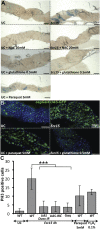
Figure 1.
Figure 1.
Immune-stimulated oxidative burst induces stem cell activation in the gut. (A) Large lacZ-marked clones containing the tubulin promoter-lacZ fusion due to mitotic recombination are observed in the guts of flies orally infected with Ecc15 or fed with paraquat. No expansion of lacZ clones was detected in the gut of flies fed with both Ecc15 and antioxidants (NAC or gluthathione). Quantification of lacZ clones is shown in Supplemental Figure S1C. (B) Ingestion of Ecc15 or paraquat induces a marked increase in the number of esgGal4, UAS-GFP-positive cells. In contrast, coingestion of Ecc15 and glutathione did not induce epithelium renewal. In unchallenged flies, most of the GFP signal corresponds to ISCs. In Ecc15-infected flies, the GFP signal was observed in both ISCs and ISC-derived daughter cells (see Supplemental Fig. S6 for distribution of escargot-positive cells along the midgut). Flies were collected 16 h post-infection. (C) Quantification of PH3-positive cells per midgut of unchallenged (UC) flies or flies collected 4 h after feeding with Ecc15, paraquat, or H2O2. Guts of Duox RNAi flies (daGal4, UAS-Duox-IR), Gαq-deficient flies, or flies overexpressing a catalase (daGal4, UAS-IRC) orally infected with Ecc15 exhibit a lower number of dividing ISCs compared with wild type. Mean ± standard deviation (SD) are shown. Values significantly different in a Student's t-test (P < 0.001) are denoted by three asterisks (***).
Figure 2.
Figure 2.
The JAK–STAT pathway is required for ISC proliferation upon Ecc15 infection. (A) Oxidative stress modulates the activation of JAK–STAT and JNK pathways upon Ecc15 infection. RT-qPCR analysis of gut extracts shows that the expression levels of JAK–STAT (upd3, Socs36E, monitored 4 h post-infection) and JNK (puckered, monitored 30 min post-infection) target genes were reduced upon Ecc15 infection in the gut of Duox RNAi flies or flies expressing a catalase (daGal4; UAS-IRC). Ingestion of paraquat was sufficient to induce upd3, Socs36E, or puckered. In contrast, the level of Diptericin (a gene tightly regulated by the Imd pathway) was not affected by conditions modulating ROS. Mean of four independent experiments ± SD are shown. (B) Quantification of PH3-positive cells per midgut shows an increase in the number of mitotic cells upon Ecc15 infection in both wild-type flies and flies with reduced JAK–STAT activity in enterocytes (NP1Gal4, UAS-STAT92E-IR), but not in ISCs (esgGal4TS, UAS-STAT92E-IR). Silencing of upd3 in enterocytes (NP1Gal4, UAS-upd3-IR), but not in ISCs (esgGal4, UAS-upd3-IR), reduced the number of mitotic ISCs in the guts of flies collected 4 h after feeding with Ecc15. Overexpression of a gain-of-function allele of hop in ISCs increases the mitotic index (esgGal4TS, UAS-hopTum). Mean ± SD are shown. Values significantly different from infected wild-type flies in a t-test at P < 0.001 are denoted by three asterisks (***). (C1–C5) A loss of stem cells was observed in the anterior region of esgGal4TS, UAS-STAT92E-IR flies raised at 29°C. In some flies, accumulation of small-nucleated escargot-positive cells were observed in the anterior part of the midgut, as indicated with a red arrow in C3. (C4) Accumulation of small-nucleated escargot-positive cells was observed in the posterior regions of the gut of all esgGal4TS, UAS-STAT92E-IR flies raised at 29°C, in the absence of infection. (C5) No increase in epithelium renewal was observed in these flies upon infection with Ecc15, as indicated by the lack of large nuclei cells expressing GFP. Expression of the esgGal4, UAS-GFP reporter gene was monitored 16 h after infection with Ecc15. (D1,D2) Overexpression of hopTum (D1) or upd3 (D2) in ISCs is sufficient to induce a high level of epithelium renewal in the absence of infection. (E) upd3 expression is induced along the gut following Ecc15 ingestion. (E1) upd3 was expressed mostly in enterocytes 4 h after Ecc15 infection, as revealed by the GFP signal (green) of upd3Gal4 UAS-GFP flies. (F) Physical damage to the gut with tweezers induced local expression of the upd3Gal4, UAS-GFP reporter within 30 min following injury.
Figure 3.
Figure 3.
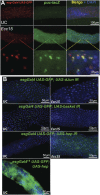
The JNK pathway is required for ISC maintenance and proliferation upon Ecc15 infection. (A) Immunostaining with antibodies against GFP (red) or LacZ (green) of esgGal4, UAS-GFP, pucE69 (puc-lacZ) flies collected 30 min after Ecc15 infection shows that the level of JNK signaling activity increases (top center panel, compared with middle center panel) upon Ecc15 infection in both enterocytes and ISCs, the staining being more intense in ISCs (merge, right panels); higher magnification is shown in the bottom panels. (B) Loss of ISCs and the absence of epithelium renewal were observed in the guts of flies with reduced JNK activity in ISCs. Guts of flies carrying the esgGal4, UAS-GFP combined with UAS-dJun-IR, UAS-basket IR, or UAS-hep-IR were examined 16 h post-infection with Ecc15. Silencing of dJun-IR in ISCs reduced the number of ISC in both unchallenged and challenged animals. In contrast, silencing of the genes encoding the JNK Basket and JNKK Hep in ISCs leads to a loss of ISCs only after infection with Ecc15. Ectopic expression of UAS-hep in ISCs induces epithelium renewal in the absence of infection.
Figure 4.
Figure 4.
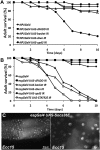
Epithelium renewal is required for proper resistance to Ecc15 infection. (A,B) Survival analysis at 29°C shows that flies impaired in epithelium renewal succumbed 4–8 d following ingestion of Ecc15. UAS-dFADD-IR, UAS-basket-IR, UAS-dJun-IR, or UAS-upd3-IR constructs were expressed in either enterocytes (NP1Gal4; A) or ISCs (esgGal4 or esgGal4TS; B). (C) Guts of flies impaired in epithelium renewal displayed altered gut morphology as revealed by the lack of DAPI nuclear staining. Flies were observed 4 d post-infection.
Figure 5.
Figure 5.
Indigenous gut microbiota activate a basal level of ISC activity. (A) Axenic (Ax) flies display a level of ISC proliferation lower than CR flies, as measured by the number of mitotic cells along the gut of unchallenged flies (PH3-positive cells). Higher numbers of dividing cells were detected in the guts of CR flies, but not axenic RelE20 or PGRP-LCE12 flies, in the absence of infection. Both young (2–4 d) and old (30 d) flies were examined. Means ± SD are shown. Means were analyzed by ANOVA and separated for significance according to Fisher's protected LSD at P = 0.05. Means designated with the same letter are not significantly different. (B) RT-qPCR analysis shows that axenic flies express lower levels of Diptericin, upd3, Socs36E, and puckered compared with CR flies in the absence of infection. Expression of these genes increased in old CR flies, but not old axenic flies. Means ± SD are shown. Values significantly different from the wild-type CR flies in a t-test at P < 0.001 are denoted for axenic wild type versus CR wild type (#), CR RelE20 versus CR wild type (≈), and old CR wild type versus young CR wild type (*). (C) Basal upd3 expression was not detected in axenic flies, as revealed by the GFP signal of upd3Gal4 UAS-GFP flies. (D1) The guts of old (30 d) CR, but not axenic, esgGal4 UAS-GFP flies display increased epithelium renewal all along the gut. (D2) Large rounded escargot-positive cells accumulate in the guts of old CR, but not axenic, wild-type flies.
Figure 6.
Figure 6.
P. entomophila (Pe) disrupts infection-induced epithelium renewal. (A) DAPI staining revealed that P. entomophila-infected guts are shorter than unchallenged guts 16 h after ingestion. The gut of P. entomophila-infected flies is composed of regions devoid of enterocytes and remaining enterocytes display abnormalities, as indicated by the loss of nuclear staining for DAPI. (B1–B8) Epithelium renewal was not observed in the gut of esgGal4, UAS-GFP flies infected with high doses of P. entomophila (OD600 > 25, B3,B4). (B3) Lack of ISCs was observed when flies were fed a high concentration of P. entomophila (OD600 = 100). (B5,B6) In contrast, infection with lower doses of P. entomophila (OD600 < 5) stimulated epithelium renewal. The avirulent P. entomophila mutant gacA induced neither epithelium renewal nor the loss of ISCs (B7), while the aprA derivative induced a slight increase of epithelium renewal (B8). Guts of esgGal4, UAS-GFP were examined 16 h after infection. (C) Infection with P. entomophila induced both JAK–STAT (Socs36E, monitored 4 h post-infection) and JNK (puckered, monitored 30 min post-infection) pathways in the gut, as revealed by RT-qPCR.
Figure 7.
Figure 7.
Modulation of gut epithelium renewal by invasive pathogens and indigenous gut microbiota. The JAK–STAT and JNK signaling pathways are required to maintain gut homeostasis upon exposure to a broad range of bacteria. In normal conditions, low levels of the indigenous gut microbiota and transient environmental microbes maintain a basal level of epithelium renewal. The increase in gut microbes in old or Imd-deficient flies is associated with a chronic activation of the JNK and JAK–STAT pathways, leading to an increase in ISC proliferation and gut disorganization. The impact of pathogenic bacteria can have different outcomes on gut homeostasis, depending on the degree of damage they inflict on the host. Damage to the gut caused by infection with E. carotovora is compensated for by an increase in epithelium renewal. Infection with a high dose of P. entomophila disrupts the homeostasis normally maintained by epithelium renewal and damage is not repaired, contributing to the death of the fly.

Comment in

Similar articles

Cited by

References

    1. Agaisse H, Petersen UM, Boutros M, Mathey-Prevot B, Perrimon N. Signaling role of hemocytes in Drosophila JAK/STAT-dependent response to septic injury. Dev Cell. 2003;5:441–450. - PubMed
    1. Amcheslavsky A, Jiang J, Ip YT. Tissue damage-induced intestinal stem cell division in Drosophila. Cell Stem Cell. 2009;4:49–61. - PMC - PubMed
    1. Bach EA, Ekas LA, Ayala-Camargo A, Flaherty MS, Lee H, Perrimon N, Baeg GH. GFP reporters detect the activation of the Drosophila JAK/STAT pathway in vivo. Gene Expr Patterns. 2007;7:323–331. - PubMed
    1. Basset A, Khush RS, Braun A, Gardan L, Boccard F, Hoffmann JA, Lemaitre B. The phytopathogenic bacteria Erwinia carotovora infects Drosophila and activates an immune response. Proc Natl Acad Sci. 2000;97:3376–3381. - PMC - PubMed
    1. Bates KL, Higley M, Letsou A. Raw mediates antagonism of AP-1 activity in Drosophila. Genetics. 2008;178:1989–2002. - PMC - PubMed

Publication types

MeSH terms