

Anti-inflammatory peptide-functionalized hydrogels for insulin-secreting cell encapsulation
- PMID: 19782393
- PMCID: PMC2784009
- DOI: 10.1016/j.biomaterials.2009.09.045
Anti-inflammatory peptide-functionalized hydrogels for insulin-secreting cell encapsulation
Abstract
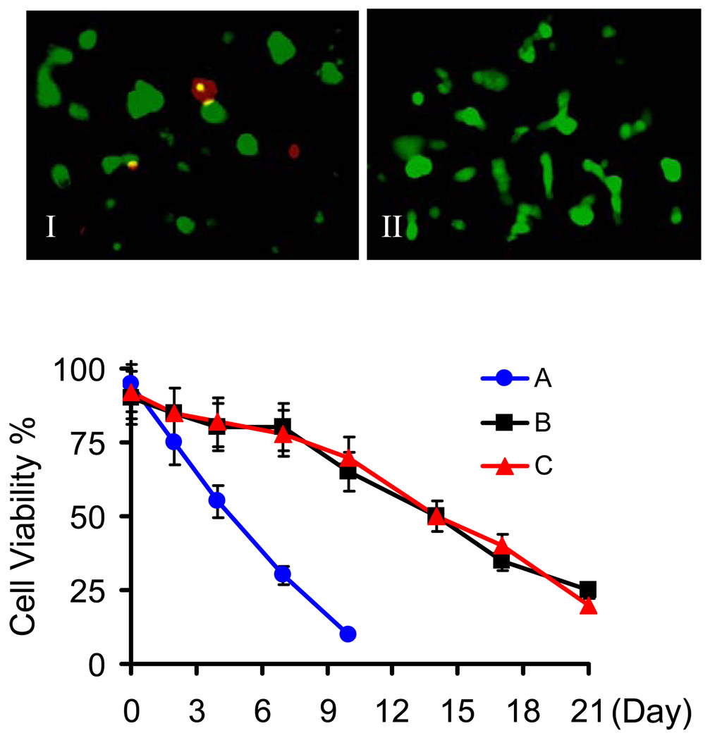
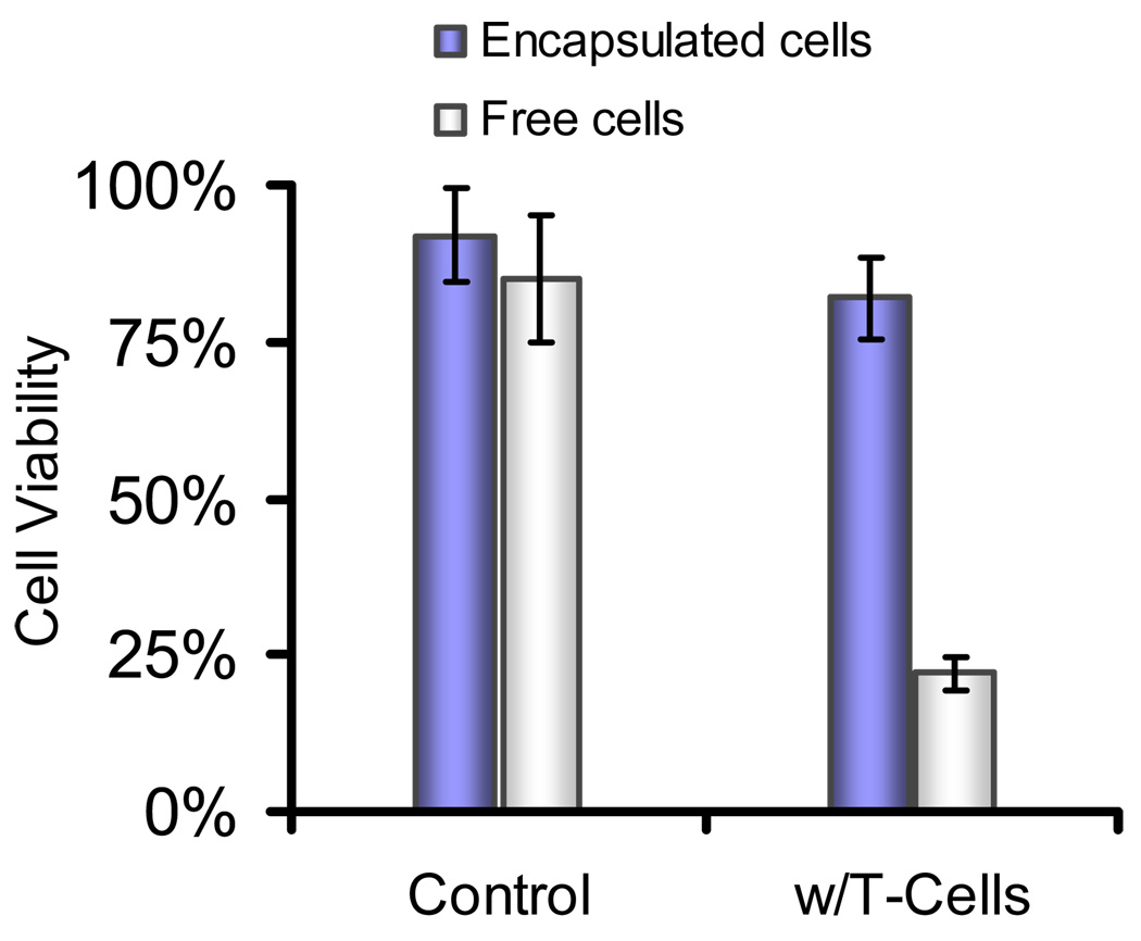
Pancreatic islet encapsulation within semi-permeable materials has been proposed for transplantation therapy of type I diabetes mellitus. Polymer hydrogel networks used for this purpose have been shown to provide protection from islet destruction by immunoreactive cells and antibodies. However, one of the fundamental deficiencies with current encapsulation methods is that the permselective barriers cannot protect islets from cytotoxic molecules of low molecular weight that are diffusible into the capsule material, which subsequently results in beta-cell destruction. Use of materials that can locally inhibit the interaction between the permeable small cytotoxic factors and islet cells may prolong the viability and function of encapsulated islet grafts. Here we report the design of anti-inflammatory hydrogels supporting islet cell survival in the presence of diffusible pro-inflammatory cytokines. We demonstrated that a poly(ethylene glycol)-containing hydrogel network, formed by native chemical ligation and presenting an inhibitory peptide for islet cell surface IL-1 receptor, was able to maintain the viability of encapsulated islet cells in the presence of a combination of cytokines including IL-1 beta, TNF-alpha, and INF-gamma. In stark contrast, cells encapsulated in unmodified hydrogels were mostly destroyed by cytokines which diffused into the capsules. At the same time, these peptide-modified hydrogels were able to efficiently protect encapsulated cells against beta-cell specific T-lymphocytes and maintain glucose-stimulated insulin release by islet cells. With further development, the approach of encapsulating cells and tissues within hydrogels presenting anti-inflammatory agents may represent a new strategy to improve cell and tissue graft function in transplantation and tissue engineering applications.
Figures


Similar articles
-
Glucagon-like peptide-1 functionalized PEG hydrogels promote survival and function of encapsulated pancreatic beta-cells.Biomacromolecules. 2009 Sep 14;10(9):2460-7. doi: 10.1021/bm900420f. Biomacromolecules. 2009. PMID: 19586041 Free PMC article.
-
The effects of cell-matrix interactions on encapsulated beta-cell function within hydrogels functionalized with matrix-derived adhesive peptides.Biomaterials. 2007 Jul;28(19):3004-11. doi: 10.1016/j.biomaterials.2007.03.005. Epub 2007 Mar 14. Biomaterials. 2007. PMID: 17391752
-
Mesenchymal stem cells and ligand incorporation in biomimetic poly(ethylene glycol) hydrogels significantly improve insulin secretion from pancreatic islets.J Tissue Eng Regen Med. 2017 Mar;11(3):694-703. doi: 10.1002/term.1965. Epub 2014 Nov 13. J Tissue Eng Regen Med. 2017. PMID: 25393526
-
Multifunctional Islet Transplantation Hydrogel Encapsulating A20 High-Expressing Islets.Drug Des Devel Ther. 2020 Sep 29;14:4021-4027. doi: 10.2147/DDDT.S273050. eCollection 2020. Drug Des Devel Ther. 2020. PMID: 33061306 Free PMC article. Review.
-
Islet Encapsulation: New Developments for the Treatment of Type 1 Diabetes.Front Immunol. 2022 Apr 14;13:869984. doi: 10.3389/fimmu.2022.869984. eCollection 2022. Front Immunol. 2022. PMID: 35493496 Free PMC article. Review.
Cited by
-
Chemical and Biological Engineering Strategies to Make and Modify Next-Generation Hydrogel Biomaterials.Matter. 2023 Dec 6;6(12):4195-4244. doi: 10.1016/j.matt.2023.10.012. Epub 2023 Nov 2. Matter. 2023. PMID: 38313360 Free PMC article.
-
Improved method for synthesis of cysteine modified hyaluronic acid for in situ hydrogel formation.Chem Commun (Camb). 2015 Jun 14;51(47):9662-5. doi: 10.1039/c5cc02367j. Chem Commun (Camb). 2015. PMID: 25977950 Free PMC article.
-
Challenges and opportunities in the islet transplantation microenvironment: a comprehensive summary of inflammatory cytokine, immune cells, and vascular endothelial cells.Front Immunol. 2023 Dec 4;14:1293762. doi: 10.3389/fimmu.2023.1293762. eCollection 2023. Front Immunol. 2023. PMID: 38111575 Free PMC article. Review.
-
Pancreatic Islet Survival and Engraftment Is Promoted by Culture on Functionalized Spider Silk Matrices.PLoS One. 2015 Jun 19;10(6):e0130169. doi: 10.1371/journal.pone.0130169. eCollection 2015. PLoS One. 2015. PMID: 26090859 Free PMC article.
-
Encapsulated islet transplantation: strategies and clinical trials.Immune Netw. 2013 Dec;13(6):235-9. doi: 10.4110/in.2013.13.6.235. Epub 2013 Dec 20. Immune Netw. 2013. PMID: 24385941 Free PMC article. Review.
References
-
- Orive G, Hernandez RM, Gascon AR, Calafiore R, Chang TMS, de Vos P, et al. History, challenges and perspectives of cell microencapsulation. Trends Biotechnol. 2004;22:87–92. - PubMed
-
- Efrat S. Cell replacement therapy for type 1 diabetes. Trends Mol Med. 2002;8:334–339. - PubMed
-
- Eftat S. Beta-cell replacement for insulin-dependent diabetes mellitus. Adv Drug Deliver Rev. 2008;60:114–123. - PubMed
-
- Gazda LS, Vinerean HV, Laramore MA, Diehl CH, Hall RD, Rubin AL, et al. Encapsulation of porcine islets permits extended culture time and insulin independence in spontaneously diabetic BB rats. Cell Transplant. 2007;16:609–620. - PubMed
-
- Wang T, Lacik I, Brissova M, Anilkumar AV, Prokop A, Hunkeler D, et al. An encapsulation system for the immunoisolation of pancreatic islets. Nat Biotechnol. 1997;15:358–362. - PubMed
Publication types
MeSH terms
Substances
Grants and funding
LinkOut - more resources
Full Text Sources
Other Literature Sources
