Optimization of differential immunogold-silver and peroxidase labeling with maintenance of ultrastructure in brain sections before plastic embedding
- PMID: 1977960
- PMCID: PMC2845158
- DOI: 10.1016/0165-0270(90)90015-8
Optimization of differential immunogold-silver and peroxidase labeling with maintenance of ultrastructure in brain sections before plastic embedding
Abstract
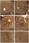
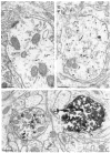
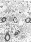
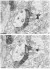
The limited success of immunogold labeling for pre-embedding immunocytochemistry of neuronal antigens is largely attributed to poor penetration of large (5-20 nm) colloidal gold particles. We examined the applicability of using silver intensification of 1 nm colloidal gold particles non-covalently bound to goat anti-rabbit immunoglobulin (1) for single labeling of a rabbit antiserum against the catecholamine synthesizing enzyme, tyrosine hydroxylase (TH), and (2) for immunogold localization of rabbit anti-TH simultaneously with immunoperoxidase labeling of a mouse monoclonal antibody against the opiate peptide, leucine-enkephalin (LE). Vibratome sections were collected from acrolein fixed brains of adult rats. These sections were immunolabeled without use of freeze-thawing or other methods that enhance penetration, but damage ultrastructure. By light microscopy, incubations in the silver intensifier (Intense M, Janssen) for less than 10 min at room temperature resulted in a brownish-red reaction product for TH. This product was virtually indistinguishable from that seen using diaminobenzidine reaction for detection of peroxidase immunoreactivity. Longer incubations produced intense black silver deposits that were more clearly distinguishable from the brown immunoperoxidase labeling. However, by light microscopy, the gold particles seen by electron microscopy were most readily distinguished from peroxidase reaction product with shorter silver intensification periods. The smaller size of gold particles with shorter periods of silver intensification also facilitated evaluation of labeling with respect to subcellular organelles. Detection of the silver product did not appear to be appreciably changed by duration of post-fixation in osmium tetroxide. In dual-labeled sections, perikarya and terminals exhibiting immunogold-silver labeling for TH were distinct from those containing immunoperoxidase labeling for LE. These results (1) define the conditions needed for optimal immunogold-silver labeling of antigens while maintaining the ultrastructural morphology in brain, and (2) establish the necessity for controlled silver intensification for light or electron microscopic differentiation of immunogold-silver and peroxidase reaction products and for optimal subcellular resolution.
Figures


Similar articles
-
Immunolabeling of retrogradely transported Fluoro-Gold: sensitivity and application to ultrastructural analysis of transmitter-specific mesolimbic circuitry.J Neurosci Methods. 1994 Nov;55(1):65-78. doi: 10.1016/0165-0270(94)90042-6. J Neurosci Methods. 1994. PMID: 7891464
-
Ultrastructural basis for interactions between central opioids and catecholamines. I. Rostral ventrolateral medulla.J Neurosci. 1989 Jun;9(6):2114-30. doi: 10.1523/JNEUROSCI.09-06-02114.1989. J Neurosci. 1989. PMID: 2566665 Free PMC article.
-
Autoradiographic detection of [125I]-secondary antiserum: a sensitive light and electron microscopic labeling method compatible with peroxidase immunocytochemistry for dual localization of neuronal antigens.J Histochem Cytochem. 1986 Jun;34(6):707-18. doi: 10.1177/34.6.2422251. J Histochem Cytochem. 1986. PMID: 2422251
-
Neuronal imaging with colloidal gold.J Microsc. 1989 Jul;155(Pt 1):27-59. doi: 10.1111/j.1365-2818.1989.tb04297.x. J Microsc. 1989. PMID: 2671381 Review.
-
Colloidal gold and biotin-avidin conjugates as ultrastructural markers for neural antigens.Q J Exp Physiol. 1984 Jan;69(1):1-33. doi: 10.1113/expphysiol.1984.sp002771. Q J Exp Physiol. 1984. PMID: 6201943 Review.
Cited by
-
Cannabinoid modulation of the dopaminergic circuitry: implications for limbic and striatal output.Prog Neuropsychopharmacol Biol Psychiatry. 2012 Jul 2;38(1):21-9. doi: 10.1016/j.pnpbp.2011.12.004. Epub 2012 Jan 11. Prog Neuropsychopharmacol Biol Psychiatry. 2012. PMID: 22265889 Free PMC article. Review.
-
Ultrastructural analysis of rat ventrolateral periaqueductal gray projections to the A5 cell group.Neuroscience. 2012 Nov 8;224:145-59. doi: 10.1016/j.neuroscience.2012.08.021. Epub 2012 Aug 20. Neuroscience. 2012. PMID: 22917613 Free PMC article.
-
Ultrastructural localization of the serotonin transporter in limbic and motor compartments of the nucleus accumbens.J Neurosci. 1999 Sep 1;19(17):7356-66. doi: 10.1523/JNEUROSCI.19-17-07356.1999. J Neurosci. 1999. PMID: 10460242 Free PMC article.
-
Kainate receptors are primarily postsynaptic to SP-containing axon terminals in the trigeminal dorsal horn.Brain Res. 2007 Dec 12;1184:149-59. doi: 10.1016/j.brainres.2007.09.070. Epub 2007 Oct 4. Brain Res. 2007. PMID: 17964552 Free PMC article.
-
Agonist-induced internalization of κ-opioid receptors in noradrenergic neurons of the rat locus coeruleus.J Chem Neuroanat. 2010 Dec;40(4):301-9. doi: 10.1016/j.jchemneu.2010.09.003. Epub 2010 Sep 25. J Chem Neuroanat. 2010. PMID: 20884346 Free PMC article.
References
-
- Anden NE, Dahlstrom A, Fuxe K, Larsson K. Further evidence for the presence of nigrostiatal dopamine neurons in the rat. Am J Anat. 1965;116:329–334. - PubMed
-
- Andre D, Vuillon Cacciuttolo G, Bosler O. GABA nerve endings in the rat red nucleus combined detection with serotonin terminals using dual immunocytochemistry. Neuroscience. 1987;23:1095–1102. - PubMed
-
- Basbaum AI. A rapid and simple silver enhancement procedure for ultrastructural localization of the retrograde tracer WGA apo HRP-Av and its use in double-label studies with post-embedding immunocytochemistry. J Histochem Cytochem. 1989;37:1811–1815. - PubMed
-
- Broadwell RD, Cataldo AM. The neuronal endoplasmic reticulum: Its cytochemistry and contribution to the endomembrane system. J Histochem Cytochem. 1983;31:1077–1088. - PubMed
-
- Cuello AC. Monoclonal antibodies in neuroanatomical research. In: Chan-Palay V, Palay SL, editors. Cytochemical Methods in Neuroanatomy. Alan R. Liss, Inc; New York, NY: 1982. pp. 151–164.
Publication types
MeSH terms
Substances
Grants and funding
LinkOut - more resources
Full Text Sources

