. 2009 Dec;39(12):3423-35.
doi: 10.1002/eji.200939441.

Novel CD8+ Treg suppress EAE by TGF-beta- and IFN-gamma-dependent mechanisms

Affiliations

Novel CD8+ Treg suppress EAE by TGF-beta- and IFN-gamma-dependent mechanisms

Mei-Ling Chen et al. Eur J Immunol. 2009 Dec.

Abstract

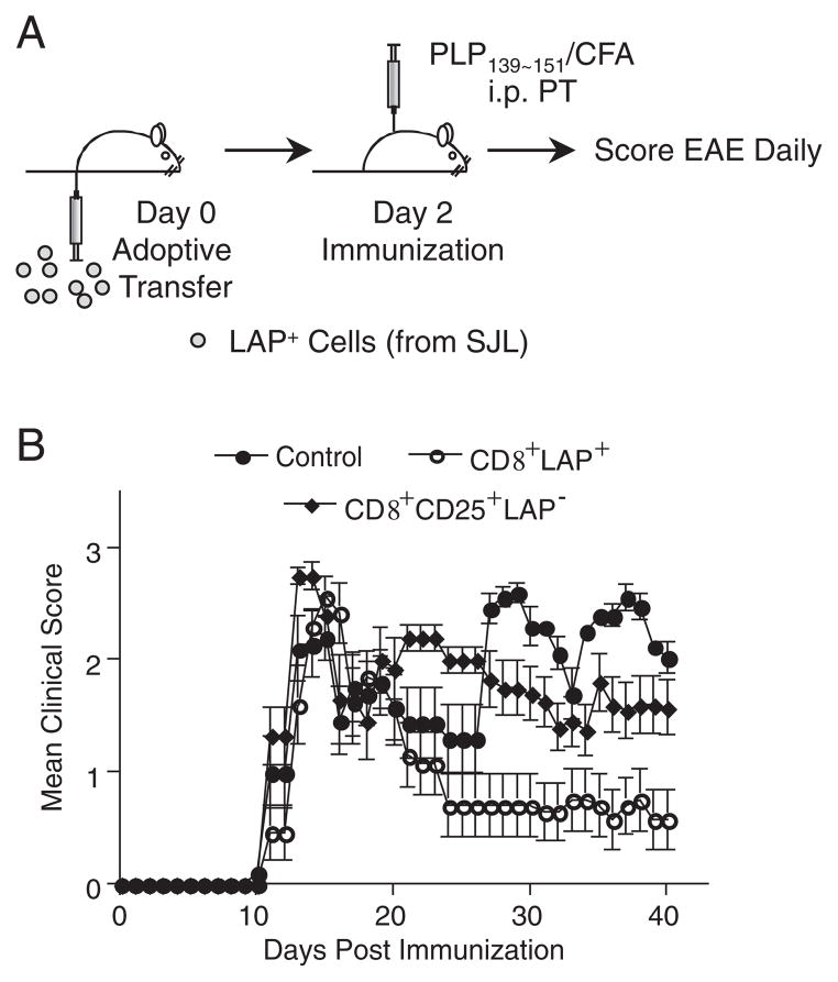
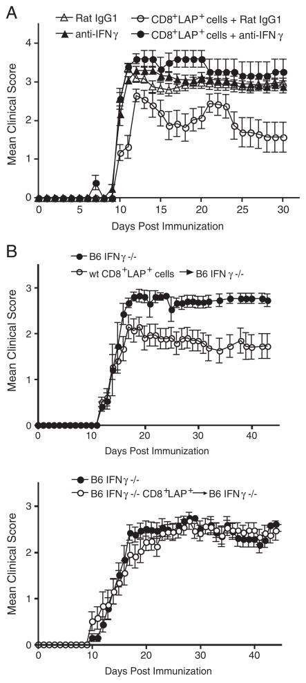
Although CD8+ Treg-mediated suppression has been described, CD8+ Treg remain poorly characterized. Here we identify a novel subset of CD8+ Treg that express latency-associated peptide (LAP) on their cell surface (CD8+LAP+ cells) and exhibit regulatory activity in vitro and in vivo. Only a small fraction of CD8+LAP+ cells express Foxp3 or CD25, although the expression levels of Foxp3 for these cells are higher than their LAP- counterparts. In addition to TGF-beta, CD8+LAP+ cells produce IFN-gamma, and these cells suppress EAE that is dependent on both TGF-beta and IFN-gamma. In an adoptive co-transfer model, CD8+LAP+ cells suppress myelin oligodendrocyte glycoprotein (MOG)-specific immune responses by inducing or expanding Foxp3+ cells and by inhibiting proliferation and IFN-gamma production in vivo. Furthermore, in vivo neutralization of IFN-gamma and studies with IFN-gamma-deficient mice demonstrate an important role for IFN-gamma production in the function of CD8+LAP+ cells. Our findings identify the underlying mechanisms that account for the immunoregulatory activity of CD8+ T cells and suggest that induction or amplification of CD8+LAP+ cells may be a therapeutic strategy to help control autoimmune processes.


Conflict of interest statement

Conflict of interest: The authors declare no financial or commercial conflict of interest.

Figures

Figure 1
Figure 1
Regulatory capacity of CD8+LAP+ cells in vitro. (A) Frequencies of CD8+LAP+ cells in splenocytes. Spleen cells obtained from naïve SJL mice were stained with CD8 and LAP-specific Ab or corresponding isotype control and analyzed by FACS. Percentage of LAP+ cells in CD8+ T cells is shown. (B) Suppressive function of CD8+LAP+ cells in vitro. 1 × 10⁵ sorted CD8+LAP+ cells purified from SJL (left panel) or B6 (right panel) mice were cultured at a 1:1 ratio with syngeneic responder CD4+CD25LAP cells. Cells were stimulated with anti-CD3 Ab (1 μg/mL) in the presence of irradiated (3000 rad) syngeneic splenic APC and assayed as described in the Materials and methods section. Data are presented as mean + SD. Percent suppression of proliferation was also shown. (C) IFN-γ production of responder CD4+CD25LAP cells in vitro. CD4+CD25LAP responder cells were cultured alone or together with CD8+LAP+ cells (ratio 1:1) in the presence of anti-CD3 Ab (1 μg/mL) and irradiated (3000 rad) syngeneic splenic APC for 60 h; IFN-γ productions by responder cells were then determined by intracellular cytokine staining. Percentage of IFN-γ+ cells among responder cells is shown. All data are representative of at least two independent experiments.
Figure 2
Figure 2
Effect of adoptive transfer of CD8+LAP+ cells on EAE. (A) Schematic representation of experimental design. 0.5 × 10⁵ sorted CD8+LAP+ or CD8+CD25+LAP cells or PBS were injected intravenously into naïve SJL mice (five mice per group). Mice were then immunized with PLP139-151 in CFA to induce EAE 2 days after adoptive transfer. (B) Mean daily score±SEM for each group (five mice per group). Data are representative of at least two independent experiments. Group that received CD8+LAP+ cells had a significant reduction in disease severity compared with control mice (p<0.001, one-way ANOVA, followed by Turkey multiple comparisons).
Figure 3
Figure 3
Cytokine profile and phenotypic characterization of CD8+LAP+ and CD8+LAP cells. (A) Cytokine production of CD8+LAP+ and CD8+LAP cells. CD8+LAP+ and CD8+LAP cells were sorted from pooled spleens and lymph nodes of naïve SJL mice as described in the Materials and methods section. After sorting, 1 × 10⁵ cells from each sorted population were stimulated with plate-bound CD3-specific Ab (10 μg/mL). Cytokines were measured by ELISA. Data show mean+SD of triplicate wells. ***p<0.0001; Student’s t-test. (B–D), Phenotypic characterization of CD8+ populations sorted by LAP expression. Flow cytometry of expression of Foxp3 (assessed by intracellular staining) (B), CD25 (C), CD45RB, and intracellular CTLA4 (D) by CD8+LAP+ and CD8+LAP cells from pooled spleens and lymph nodes of naïve SJL mice. Numbers next to outlined areas in (B) and (C) indicate percent cells positive for marker among each population. Red line in (D): marker specific staining for CD8+LAP cells; blue line: marker specific staining for CD8+LAP+ cells. All data are representative of three independent experiments.
Figure 4
Figure 4
Effect of adoptive co-transfer of CD8+LAP+ cells on the effector function of MOG TCR Tg T cells in vivo. (A) Schematic representation of experimental design. MOG TCR Tg Thy1.1+ T cells depleted of CD25+ cells, CFSE-labeled (3 × 10⁵) were transferred alone or together with CD8+ LAP+ cells into B6 (Thy1.2+) mice (three mice per group). Two days later, recipients were immunized with 50 μg of MOG35–55 peptide in CFA. Mice were killed 5 days after immunization, and cells from draining lymph nodes were harvested, stained, and analyzed by flow cytometry. (B) Proliferation of adoptively transferred MOG TCR Tg T cells (CD4+Thy1.1+) in the draining lymph nodes after immunization. The plots show the expression of CD4 versus CFSE fluorescence intensity on gated donor-derived cells (CD4+Thy1.1+). Numbers above bracketed areas indicate the frequency of CFSE+ cells among CD4+Thy1.1+ cells. p =0.015 (Student’s t-test). (C) IFN-γ production of transferred MOG TCR Tg T cells. Draining lymph node cells from mice adoptively transferred and immunized as described in (A) were restimulated ex vivo with PMA/ionomycin and stained for intracellular IFN-γ. Numbers next to bracketed areas indicate the frequency of IFN-γ+ cells among CD4+Thy1.1+ cells. p =0.015 (Student’s t-test). (D) Expression of Foxp3 of transferred MOG TCR Tg T cells. Draining lymph node cells from mice adoptively transferred and immunized as described in (A) were stained for intracellular Foxp3. Numbers next to bracketed areas indicate the frequency of Foxp3+ cells among CD4+Thy1.1+ cells. p =0.04 (Student’s t-test). (B–D) Data show mean±SD, n =3, and are representative of three independent experiments.
Figure 5
Figure 5
TGF-β-mediated suppressive function of CD8+LAP+cells in vitro and in vivo. (A) Effect of inhibitor of TGF-β signaling (ALK5 inhibitor II) on the in vitro suppressive function of CD8+LAP+ cells. CD4+CD25LAP responder cells were cultured alone or together with CD8+LAP+ at a 1:1 ratio and stimulated with anti-CD3 Ab (1 μg/mL) and irradiated (3000 rad) syngeneic splenic APC in the presence of indicated concentrations of ALK5 inhibitor and assayed as described in the Materials and methods section. Data are representative of at least two independent experiments and are presented as means±SD. Percent suppression of proliferation was shown. (B) Effect of neutralization of TGF-β on the regulatory function of CD8+LAP+ cells in vivo. SJL mice were adoptively transferred with 0.5 × 10⁵ sorted CD8+LAP+ cells 2 days before EAE induction. Mice then received five intraperitoneal injections of 50 μg of neutralizing TGF-β specific or control Ab every other day starting 1 day before EAE induction. The mean daily score±SEM for each group (five mice per group) is shown. Data are representative of at least two independent experiments. In vivo administration of anti-TGF-β significantly reversed the suppression of EAE mediated by CD8+ LAP+ cells; p<0.001 (one-way ANOVA, followed by Turkey multiple comparisons) as compared between CD8+LAP+ cells+anti-TGF-β and CD8+LAP+ cells+Rat IgG1 groups.
Figure 6
Figure 6
IFN-γ-mediated suppressive function of CD8+LAP+cells in vitro. Effect of IFN-γ and IFN-γ receptor deficiency on the in vitro suppressive function of CD8+LAP+ cells. Responder CD4+CD25LAP cells from WT or IFN-γR−/− mice were cultured alone or at 1:1 ratio with CD8+LAP+ cells from WT B6 mice (A), IFN-γR−/− mice (B); (C) responder from WT mice were cultured alone or at 1:1 ratio with CD8+LAP+ cells from WT B6 mice or IFN-γ−/− mice. In vitro proliferation assays were performed as described in the Materials and methods section. Data show mean+SD of triplicate wells and are representative of two independent experiments. Percent suppression of proliferation is also shown.
Figure 7
Figure 7
IFN-γ-mediated suppressive function of CD8+LAP+ cells in vivo. (A) Effect of neutralization of IFN-γ on the regulatory function of CD8+LAP+ cells in vivo. SJL mice were adoptively transferred with 0.5 × 10⁵ sorted CD8+LAP+ cells 2 days before EAE induction. Mice then received five intraperitoneal injections of 50 μg of neutralizing IFN-γ specific or control Ab every 3 days starting 1 day before EAE induction. Mean daily score±SEM for each group (five mice per group). Data are representative of at least two independent experiments. p<0.001 (one-way ANOVA, followed by Turkey multiple comparisons) as compared between CD8+LAP+ cells+Rat IgG1 and CD8+LAP+ cells+anti-IFN-γ groups. (B) Effect of IFN-γ deficiency on the regulatory function of CD8+LAP+ cells in vivo. 1.7 × 10⁵ sorted CD8+LAP+ cells from WT or IFN-γ−/− mice were injected intravenously into naïve IFN-γ−/− mice. Mice were then immunized with MOG35–55 in CFA to induce EAE 2 days after adoptive transfer. Mean daily score±SEM for each group (five to six mice per group). Data are representative of three independent experiments. Group that received CD8+LAP+ cells purified from WT mice had a significant reduction in disease severity compared with control mice (p<0.0001, Mann–Whitney U test).

Similar articles

Cited by

References

    1. Sakaguchi S. Naturally arising CD4+ regulatory T cells for immunologic self-tolerance and negative control of immune responses. Annu Rev Immunol. 2004;22:531–562. - PubMed
    1. Shevach EM. Regulatory T cells in autoimmmunity. Annu Rev Immunol. 2000;18:423–449. - PubMed
    1. Miyara M, Sakaguchi S. Natural regulatory T cells: mechanisms of suppression. Trends Mol Med. 2007;13:108–116. - PubMed
    1. Friese MA, Fugger L. Autoreactive CD8+ T cells in multiple sclerosis: a new target for therapy? Brain. 2005;128:1747–1763. - PubMed
    1. Jiang H, Zhang SI, Pernis B. Role of CD8+ T cells in murine experimental allergic encephalomyelitis. Science. 1992;256:1213–1215. - PubMed

Publication types

MeSH terms

LinkOut - more resources