. 2009 Oct;10(10):1439-57.
doi: 10.1111/j.1600-0854.2009.00967.x.

HCMV-encoded glycoprotein M (UL100) interacts with Rab11 effector protein FIP4

Affiliations

HCMV-encoded glycoprotein M (UL100) interacts with Rab11 effector protein FIP4

Magdalena A Krzyzaniak et al. Traffic. 2009 Oct.

Abstract

The envelope of human cytomegalovirus (HCMV) consists of a large number of glycoproteins. The most abundant glycoprotein in the HCMV envelope is the glycoprotein M (UL100), which together with glycoprotein N (UL73) form the gM/gN protein complex. Using yeast two-hybrid screening, we found that the gM carboxy-terminal cytoplasmic tail (gM-CT) interacts with FIP4, a Rab11-GTPase effector protein. Depletion of FIP4 expression in HCMV-infected cells resulted in a decrease in infectious virus production that was also associated with an alteration of the HCMV assembly compartment (AC) phenotype. A similar phenotype was also observed in HCMV-infected cells that expressed dominant negative Rab11(S25N). Recently, it has been shown that FIP4 interactions with Rab11 and additionally with Arf6/Arf5 are important for the vesicular transport of proteins in the endosomal recycling compartment (ERC) and during cytokinesis. Surprisingly, FIP4 interaction with gM-CT limited binding of FIP4 with Arf5/Arf6; however, FIP4 interaction with gM-CT did not prevent recruitment of Rab11 into the ternary complex. These data argued for a contribution of the ERC during cytoplasmic envelopment of HCMV and showed a novel FIP4 function independent of Arf5 or Arf6 activity.


Figures

Figure 1
Figure 1. The gM cytoplasmic tail (gM-CT) interacts with FIP4
(A) Diagram representing gM protein fragment (300–372aa) which was cloned and used as a bait during yeast two hybrid screening and N-terminal GST tagged constructs of gM-CT: gM-ac1 and gM-ac2 which were used for the pull down assays of FIP4-myc. (B) Diagram of the FIP4 protein with depicted domains: N-terminal EF-hand, CC coil-coiled domain responsible for the FIP4 homo- and heterodimerization with FIP3, Rab Binding Domain (RBD) which interacts with Rab11, 454-543 aa region which is essential for the interaction with Arf5/Arf6 and was recovered as a cDNA clone of gM-CT interactions during yeast two hybrid screening.
Figure 2
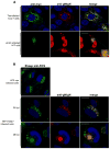
Figure 2. Immunofluorescence co-localization of FIP4 and gM/gN complex in transfected and HCMV infected cells
(A) Cos7 cells were co-transfected with constructs expressing FIP4-myc, gM and gN, 48 hours post transfection cells were fixed and reacted with anti-myc and anti-gM/gN mab followed with FITC and TRTC labeled secondary antibody as described in Material and Methods (top panel). HFF cells were electroporated with FIP4-myc construct and then infected with HCMV, 6 dpi cells were fixed and reacted with anti-myc, anti-gM/gN and anti-IE1 mab (detecting immediate early antigen-1 expressed in the nucleus of the infected cells) antibody and appropriate secondary antibody (bottom panel). (B) To detect co-localization of endogenous FIP4 in HCMV infected cells with gM/gN complex. HFF cells were infected with HCMV, 24 and 48 hpi cells were fixed and reacted with a polyclonal anti-FIP4 sheep antibodies and anti-gM/gN mab and appropriate secondary antibody. Note that before staining cells were reacted with anti-FC receptor antibody to block non-specific FC receptor staining (data not shown). In addition, non-immune goat antibodies did not stain infected cells (data not shown). Bar in all merged images indicates 20 μm.
Figure 3
Figure 3. FIP4 interact with the gM-CT
(A) Pull down assay in which glutathione sepharose beads containing gM-CT constructs (GST-gM-CT, GST-gM-ac1, GST-gM-ac2) or purified GST alone were incubated with the HK293 cell lysate expressing FIP4-myc. After extensive washing protein samples were boiled in sample buffer, resolved by SDS-PAGE and assayed by western analysis. Western blots were probed with anti-myc (9E10) mab and detected with the HRP (top panel). The lower panel shows the Coomassie blue stained gels of the GST purified input of proteins used in pull down assay. (B) Pull down assay of FIP3 or FIP4 by cytoplasmic tail of gM. Glutathione sepharose beads containing gM-CT constructs or GST alone were incubated with HK293 cell lysates expressing FIP3-myc or FIP4-myc. After extensive washing, beads were boiled and eluted proteins resolved by SDS-PAGE followed by western blot analysis. Western blots were probed with anti-myc mab and detected with the HRP. (C) Fluorescence Resonance Energy Transfer (FRET) indicating strong FIP4 and gM interaction in HCMV infected cells. For FRET assays, HFF cells were electroporated with FIP4 or FIP3 myc-tagged constructs and plated on the coverslips. 24 hours later, the cells were infected with HCMV. The cells were fixed in 4% PFA and stained with IMP anti-gM monoclonal antibodies (specific for the C-terminal tail of gM) or anti-myc antibody followed by labeling with secondary antibody conjugated with FITC or TxRed. A non-bleaching region was selected as an internal control of FRET analysis detail description of the assay is provided in Materials and Methods.
Figure 4
Figure 4. shRNA depletion of the FIP4 expression
(A) HK293 cells were co-transfected with FIP4-myc together with either shRNA to deplete FIP4 expression or with scrambled control shRNA. The protein samples were collected 2, 3, and 4 days post transfection and frozen in −80°C. The control cell sample transfected only with FIP4-myc was harvested on the day 4 post transfection. All cell pellets were lysed in equal amounts of sample buffer and then resolved by SDS-PAGE, followed by analysis by western blotting. Western blots were probed with anti-myc to detect expression of the FIP4-myc (top panel). Additionally, the membranes were probed with anti-actin antibody as a loading control (bottom panel). The histogram represents efficiency of shRNA transfection in HK293 cells. The percentage of transfected cells was calculated as number of GFP expressing cells, 2 and 4 days post transfection per 100 cells counted stained with DAPI as described in Materials and Methods. (B) HFF cells were electroporated using Amaxa with shRNA to deplete FIP4 expression or with scrambled control shRNA and the cells were plated on 13 mm coverslips. On day 4 and 6 post electroporation cells were fixed and stained with DAPI to visualized total number of cells on the cover slips. The efficiency of shRNA expression was estimated by calculating number of GFP expressing cells per 100 cells stained nuclei as described in the Materials and Methods. HFF cells were electroporated with FIP4-myc and either with shRNA mix to deplete FIP4 or a control scrambled shRNA. One day post electroporation, cells were infected with AD169 HCMV. On 2, 4, and 6 days post infection HFF cells were harvested in lysis buffer, subjected to SDS-PAGE and analyzed by western blotting. FIP-4 expression was detected with anti-myc mab (top panel). The membrane was stripped and than probed with anti-p115 (middle panel) and anti-UL85 mab (bottom panel) as an off target control for the shRNA. (C) HFF cells were electroporated with FIP3-myc and the FIP4 shRNA mix or control scrambled shRNA. One day post electroporation cells were infected with AD169 HCMV. On 2, 4, and 6 days post infection HFF cells were harvested in lysis buffer, resolved by SDS-PAGE gel, and analyzed by western blotting. FIP3 was detected with anti-myc mab.
Figure 5
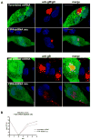
Figure 5. Depletion of the FIP4 expression alters the assembly compartment formation and leads to decrease in HCMV virus production
(A-B) HFF cells were electroporated with shRNA to deplete FIP4 or with control scrambled shRNA sequence and plated on 13 mm glass coverslips. On the following day, the cells were infected with HCMV at the multiplicity of infection (moi) of 1.0 and 4 days later cells were fixed and stained with anti-gM/gN mab (A) or anti-gB mab (B) as described in Materials and Methods. Note that all images were collected with the identical laser intensity and gain settings, thus allowing for comparison of signal intensity. The assembly compartment is not well formed in cells depleted FIP4 expression by shRNA. Bar in all merged images indicates 20 μm. (C) HFF expressing shRNA vectors to deplete FIP4 or control scrambled shRNA were infected with AD169 at an moi of 0.1 and total cultures (supernatant and cells) in 1.5 ml media were harvested at the indicated time points post infection. Cell supernatant and disrupted cells were combined and assayed for the amount of the infectious particles using a fluorescence based infectivity assay as described in Materials and Methods. Results are expressed as a log10 infectious units/ml of sample.
Figure 6
Figure 6
FIP4 bound to the gM-CT recruits Rab11 but fails to bind Arf5 or Arf6. HK293 cells were transfected with FIP4-myc alone or co-transfected with FIP4-myc and either with HA-Arf5, HA-Arf6, GFP-Rab11, and Arf1-HA as a control. Two days post transfection cells were lysed and analyzed by immunoprecipitation with myc antibody-tagged magnetic beads as described in the Materials and Methods or incubated with sepharose beads containing purified GST-gM-CT or GST alone. After immunoprecipitation and extensive washing, precipitated protein were resolved by SDS-PAGE, transferred to membranes and probed with appropriate antibody. The asterisk indicates HA-Arf6 band which was visualized only after prolonged exposure of the membrane that had been probed with anti-HA and developed with HRP.
Figure 7
Figure 7. Expression of the dominant negative HA-Arf6(T27N) does not limit HCMV virus assembly and production
(A) Analysis of gM/gN localization in the HCMV infected cells expressing HA-Arf6 or dominant negative HA-Arf6 (T27N). HFF cells were electroporated with plasmids encoding HA-Arf6 or HA-Arf6 (T27N) and plated on 13 mm glass coverslips. Twenty-four hours later cells were infected with HCMV at an moi of 1.0 then 4 days post infection (dpi) cells were fixed and stained with anti-gM/gN and with anti-HA antibody, as described in the Methods. Bar in all merged images indicates 20 μm. (B) HCMV growth kinetics in HFF cells expressing HA-Arf6 or dominant negative HA-Arf6 (T27N). HFF expressing HA-Arf6 or HA-Arf6 (T27N) were infected with HCMV at a moi of 0.1 and total cultures (supernatant and cells) in 1.5 ml media were harvested at the indicated time points post infection. Cell supernatant and disrupted cells were combined and assayed for the amount of the infectious particles using a fluorescence based infectivity assay described in the Materials and Methods. Results were expressed as a log10 infectious units/ml of sample.
Figure 8
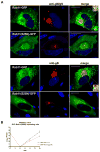
Figure 8. Expression of the dominant negative GFP-Rab11(S25N) alters formation of the assembly compartment and leads to a decrease in HCMV virus yield
(A–B) HFF cells were electroporated with GFP-Rab11 or dominant negative GFP-Rab11(S25N) and plated on 13 mm glass coverslips. On the following day, cells were infected with HCMV at an moi of 1.0 and 4 dpi cells were fixed and stained with anti-gM/gN (14-16A) (A) or anti-gB mab (B), as described in the Material and Methods. Note that all images were collected with the identical laser intensity and gain. In cells expressing dominant negative GFP-Rab11(S25N) visualized by the GFP expression there was a decrease in the signal from the gM/gN complex as well as gB within the AC as compared to the cells that did not express the dominant negative Rab11(S25N). Bar in all merged images indicates 20 μm. (C) HCMV growth kinetics in HFF cells expressing Rab11 or GFP-Rab11(S25N). HFF expressing GFP constructs of Rab11 and Rab11(S25N) were infected with HCMV at an moi of 0.1 and total cultures (supernatant and cells) in 1.5 ml media were harvested at the indicated time points post infection. Cell supernatant and disrupted cells were combined and assayed for the amount of the infectious particles using a fluorescence based infectivity assay described in the Materials and Methods. Results were expressed as a log10 infectious units/ml of sample.
Figure 9
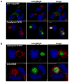
Figure 9. Assembly compartment formation in HFF cells is associated with ERC markers and does not contain markers of the late endocytic pathway
(A) HFF cells were infected with HCMV and on day 6 post infection, cells were washed extensively in serum free media and incubated for two hours with the EGF-Alexa488 or transferrin (Tf)-TRITC in serum free media. After washing cells were fixed and stained with anti-gM/gN mab followed with anti-IgM FITC or TRITC respectively. Cells were imaged by confocal microscopy. Bar in all merged images indicates 20 μm. (B) HFF were electroporated with RFP-tagged cathepsin-D, infected with HCMV, and on day 6 post infection, cells were stained with anti-gM/gN mab (top panel). Alternatively, on day 6 following infection with HCMV, HFF cells were stained with anti-CD63 and anti-gM/gN and developed with TRTC goat-anti-mouse IgG1 or FITC goat anti-mouse IgM respectively (bottom panel). All images were collected at 100× magnification.
Figure 10
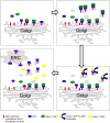
Figure 10. Model of FIP4 function and gM/gN trafficking to the AC of HCMV infected HFF cells
(A) gM/gN complex transits through secretory pathway after reaching TGN, the gM cytoplasmic tail interacts with FIP4. (B) Fip4 interacting with gM recruits Rab11-GTP and facilitates export from TGN to ERC. (C) Vesicles exported from TGN induces Rab11 GTP-GDP hydrolysis on gM/gN-FIP4 coated vesicles and Rab11-GDP dissociation from the vesicles. (D) gM-FIP4 containing vesicles reach ERC. Progressive accumulation of the gM/gN in the ERC and possibly other glycoproteins leads to the formation of the AC a cellular compartment containing host cell proteins of the ERC.

Similar articles

Cited by

References

    1. Yoshikawa T. Significance of human herpesviruses to transplant recipients. Curr Opin Infect Dis. 2003 Dec;16(6):601–6. - PubMed
    1. Halwachs-Baumann G. Recent developments in human cytomegalovirus diagnosis. Expert Rev Anti Infect Ther. 2007 Jun;5(3):427–39. - PubMed
    1. Mocarski ES, Courcelle CT. Cytomegaloviruses and their replication. 4. Philadelphia, Pa: Lippincott Williams & Wilkins; 2001.
    1. Trus BL, Gibson W, Cheng N, Steven AC. Capsid structure of simian cytomegalovirus from cryoelectron microscopy: evidence for tegument attachment sites. J Virol. 1999 Mar;73(3):2181–92. - PMC - PubMed
    1. Yu X, Shah S, Atanasov I, Lo P, Liu F, Britt WJ, et al. Three-dimensional localization of the smallest capsid protein in the human cytomegalovirus capsid. J Virol. 2005 Jan;79(2):1327–32. - PMC - PubMed

Publication types

MeSH terms

LinkOut - more resources