

Cerebellum of the premature infant: rapidly developing, vulnerable, clinically important
- PMID: 19745085
- PMCID: PMC2799249
- DOI: 10.1177/0883073809338067
Cerebellum of the premature infant: rapidly developing, vulnerable, clinically important
Abstract
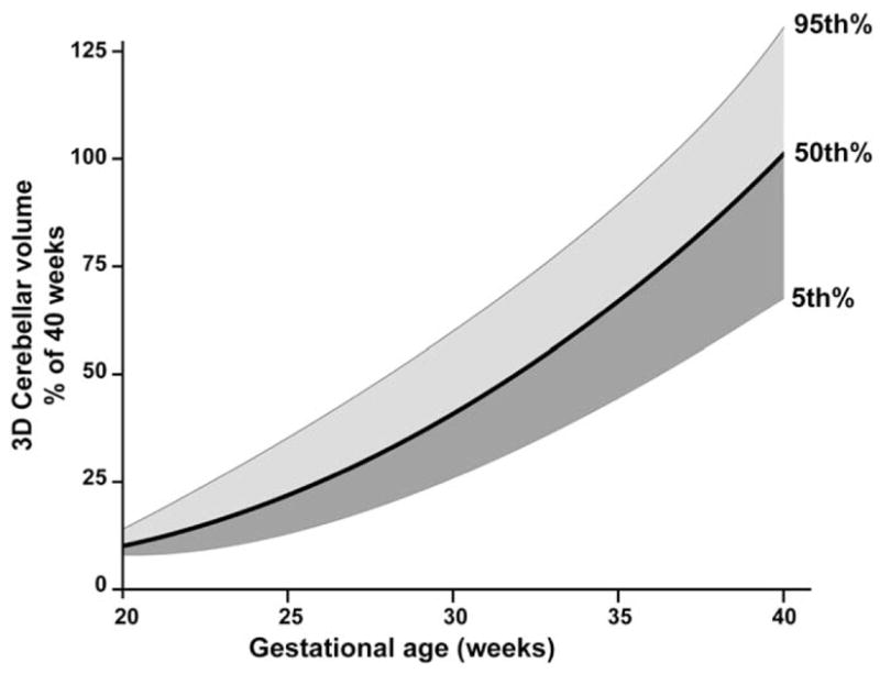
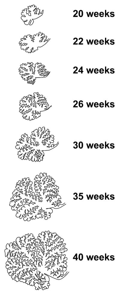
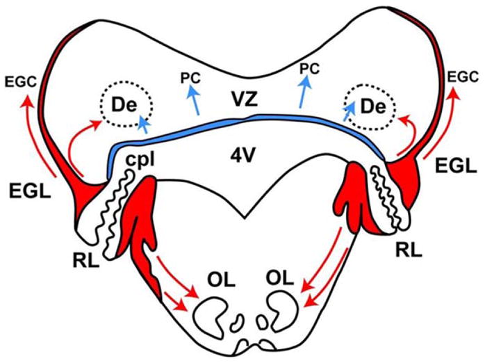
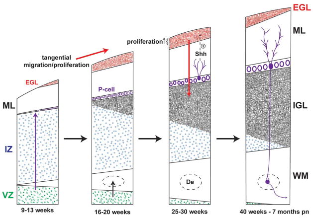
Brain abnormality in surviving premature infants is associated with an enormous amount of neurodevelopmental disability, manifested principally by cognitive, behavioral, attentional, and socialization deficits, most commonly with only relatively modest motor deficits. The most recognized contributing neuropathology is cerebral white matter injury. The thesis of this review is that acquired cerebellar abnormality is a relatively less recognized but likely important cause of neurodevelopmental disability in small premature infants. The cerebellar disease may be primarily destructive (eg, hemorrhage, infarction) or primarily underdevelopment. The latter appears to be especially common and relates to a particular vulnerability of the cerebellum of the small premature infant. Central to this vulnerability are the extraordinarily rapid and complex developmental events occurring in the cerebellum. The disturbance of development can be caused either by direct adverse effects on the cerebellum, especially the distinctive transient external granular layer, or by indirect remote trans-synaptic effects. This review describes the fascinating details of cerebellar development, with an emphasis on events in the premature period, the major types of cerebellar abnormality acquired during the premature period, their likely mechanisms of occurrence, and new insights into the relation of cerebellar disease in early life to subsequent cognitive/behavioral/attentional/socialization deficits.
Conflict of interest statement
The authors have no conflicts of interest to disclose with regard to this article.
Figures


Similar articles
-
Preterm birth and cerebellar neuropathology.Semin Fetal Neonatal Med. 2016 Oct;21(5):305-11. doi: 10.1016/j.siny.2016.04.006. Epub 2016 May 5. Semin Fetal Neonatal Med. 2016. PMID: 27161081 Review.
-
Cerebellar hemorrhagic injury in premature infants occurs during a vulnerable developmental period and is associated with wider neuropathology.Acta Neuropathol Commun. 2013 Oct 21;1:69. doi: 10.1186/2051-5960-1-69. Acta Neuropathol Commun. 2013. PMID: 24252570 Free PMC article.
-
Impaired trophic interactions between the cerebellum and the cerebrum among preterm infants.Pediatrics. 2005 Oct;116(4):844-50. doi: 10.1542/peds.2004-2282. Pediatrics. 2005. PMID: 16199692
-
Cerebellar injury in preterm infants.Handb Clin Neurol. 2018;155:49-59. doi: 10.1016/B978-0-444-64189-2.00003-2. Handb Clin Neurol. 2018. PMID: 29891076 Review.
-
Cerebellar hypoplasia of prematurity: Causes and consequences.Handb Clin Neurol. 2019;162:201-216. doi: 10.1016/B978-0-444-64029-1.00009-6. Handb Clin Neurol. 2019. PMID: 31324311 Review.
Cited by
-
Antenatal Exposure to Magnesium Sulfate Is Associated with Reduced Cerebellar Hemorrhage in Preterm Newborns.J Pediatr. 2016 Nov;178:68-74. doi: 10.1016/j.jpeds.2016.06.053. Epub 2016 Jul 22. J Pediatr. 2016. PMID: 27453378 Free PMC article.
-
Early Diagnostics and Early Intervention in Neurodevelopmental Disorders-Age-Dependent Challenges and Opportunities.J Clin Med. 2021 Feb 19;10(4):861. doi: 10.3390/jcm10040861. J Clin Med. 2021. PMID: 33669727 Free PMC article. Review.
-
Epigenetic and genetic variation at the IGF2/H19 imprinting control region on 11p15.5 is associated with cerebellum weight.Epigenetics. 2012 Feb;7(2):155-63. doi: 10.4161/epi.7.2.18910. Epigenetics. 2012. PMID: 22395465 Free PMC article.
-
Oligodendroglial maldevelopment in the cerebellum after postnatal hyperoxia and its prevention by minocycline.Glia. 2015 Oct;63(10):1825-39. doi: 10.1002/glia.22847. Epub 2015 May 12. Glia. 2015. PMID: 25964099 Free PMC article.
-
Spatiotemporal expansion of primary progenitor zones in the developing human cerebellum.Science. 2019 Oct 25;366(6464):454-460. doi: 10.1126/science.aax7526. Epub 2019 Oct 17. Science. 2019. PMID: 31624095 Free PMC article.
References
-
- Woodward LJ, Edgin JO, Thompson D, Inder TE. Object working memory deficits predicted by early brain injury and development in the preterm infant. Brain. 2005;128(pt 11):2578–2587. - PubMed
-
- Bayless S, Stevenson J. Executive functions in school-age children born very prematurely. Early Hum Dev. 2007;83(4):247–254. - PubMed
-
- Platt MJ, Cans C, Johnson A, et al. Trends in cerebral palsy among infants of very low birthweight (<1500 g) or born prematurely (<32 weeks) in 16 European centres: a database study. Lancet. 2007;369(9555):43–50. - PubMed
-
- Larroque B, Ancel PY, Marret S, et al. Neurodevelopmental disabilities and special care of 5-year-old children born before 33 weeks of gestation (the EPIPAGE study): a longitudinal cohort study. Lancet. 2008;371(9615):813–820. - PubMed
-
- Kobaly K, Schluchter M, Minich N, et al. Outcomes of extremely low birth weight (<1 kg) and extremely low gestational age (<28 weeks) infants with bronchopulmonary dysplasia: effects of practice changes in 2000 to 2003. Pediatrics. 2008;121(1):73–81. - PubMed
Publication types
MeSH terms
Grants and funding
LinkOut - more resources
Full Text Sources
Medical
