. 2009 Sep;128(1 Suppl):e837-48.
doi: 10.1111/j.1365-2567.2009.03093.x. Epub 2009 Mar 23.

Modulation of dendritic cell maturation and function with mono- and bifunctional small interfering RNAs targeting indoleamine 2,3-dioxygenase

Affiliations

Modulation of dendritic cell maturation and function with mono- and bifunctional small interfering RNAs targeting indoleamine 2,3-dioxygenase

Gro F Flatekval et al. Immunology. 2009 Sep.

Abstract

Antigen-presenting cells expressing indoleamine 2,3-dioxygenase (IDO) play a critical role in maintaining peripheral tolerance. Strategies to inhibit IDO gene expression and enhance antigen-presenting cell function might improve anti-tumour immunity. Here we have designed highly effective anti-IDO small interfering (si) RNAs that function at low concentrations. When delivered to human primary immune cells such as monocytes and dendritic cells (DCs), they totally inhibited IDO gene expression without impairing DC maturation and function. Depending on the design and chemical modifications, we show that it is possible to design either monofunctional siRNAs devoid of immunostimulation or bifunctional siRNAs with gene silencing and immunostimulatory activities. The latter are able to knockdown IDO expression and induce cytokine production through either endosomal Toll-like receptor 7/8 or cytoplasmic retinoid acid-inducible gene 1 helicase. Inhibition of IDO expression with both classes of siRNAs inhibited DC immunosuppressive function on T-cell proliferation. Immature monocyte-derived DCs that had been transfected with siRNA-bearing 5'-triphosphate activated T cells, indicating that, even in the absence of external stimuli such as tumour necrosis factor-alpha, those DCs were sufficiently mature to initiate T-cell activation. Collectively, our data highlight the potential therapeutic applications of this new generation of siRNAs in immunotherapy.


Figures

Figure 1
Figure 1
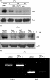
Silencing potency of indoleamine 2,3-dioxygenase (IDO) small interfering (si) RNAs in human monocytes. (a) Down-regulation of IDO gene expression by siRNAs. Freshly isolated monocytes were transfected with anti-IDO siRNAs (3 μg/225 pmol each) or control siRNA targeting β-galatosidase (3 μg/225 pmol) using nucleofection. Subsequently, IDO expression was induced with interferon-γ (IFN-γ; 500 U/ml) for 18 hr and IDO protein levels were determined by Western blots. (b) Concentration-dependent down-regulation of IDO gene expression by siRNAs. Freshly isolated monocytes were transfected with various concentrations of IDO siRNAs and then processed as in (a). (c) Quantification of the IDO signals shown in (b). The control intensity is defined as 100%. (d) Anti-IDO siRNAs are specific. Monocytes were transfected with the indicated siRNA duplexes (1 μg/75 pmol) and processed as in (a). Sca = a scrambled IDO siRNA-4. Not transfected but IFN-γ-stimulated cells were included in these experiments.
Figure 2
Figure 2
Silencing potency of indoleamine 2,3-dioxygenase (IDO) small interfering (si) RNAs in human dendritic cells (DCs). (a) Down-regulation of IDO gene expression by siRNAs in immature monocyte-derived (immo) DCs. To generate immoDCs, monocytes were treated with granulocyte–macrophage colony-stimulating factor (GM-CSF; 25 ng/ml) and interleukin-4 (IL-4; 50 ng/ml) for 5 days. Then the cells were transfected with various concentrations of IDO siRNA-3 (0·1–1·5 μg/7·5–112·5 pmol) or control siRNA (1·5 μg/112·5 pmol). Subsequently, IDO gene expression was induced by interferon-γ (IFN-γ; 500 U/ml) for 18 hr and IDO protein levels were determined by Western blots. (b) Down-regulation of IDO gene expression by siRNAs in mature monocyte-derived (mmo) DCs. To generate mmoDCs, immoDCs were stimulated with tumour necrosis factor-α (TNF-α; 50 ng/ml) for 2 days, and then they were washed, transfected with various IDO siRNAs (0·1 μg/7·5 pmol each) and processed as in (a). (c) Reverse transcription–polymerase chain reaction (RT-PCR) analysis of IDO mRNA levels. ImmoDCs were transfected with siRNA-3 (0·4 μg/30 pmol) and then IDO expression was induced with IFN-γ for 18 hr. Subsequently, total RNA was prepared and IDO and β-actin messenger RNA levels were analysed by RT-PCR as described in Materials and methods.
Figure 3
Figure 3
Efficient small interfering (si) RNA gene silencing in dendritic cells (DCs) using the BTX electroporation method. (a) Transfection of indoleamine 2,3-dioxygenase (IDO) siRNAs with BTX electroporation in immature monocyte-derived (immo) DCs. Around 5 × 10⁶ cells were transfected with IDO siRNA-4 (0·5 μg/37·5 pmol) or control siRNA (0·5 μg/37·5 pmol) and then IDO expression was induced with interferon-γ (IFN-γ; 500 U/ml) for 18 hr. Subsequently, IDO and actin protein levels were determined by Western blots. (b) IDO siRNA remains functional during DC maturation. The immoDCs were transfected with IDO siRNA- 3 (1·5 μg/112·5 pmol) or control siRNA (1·5 μg/112·5 pmol) and then the cells were cultured in the presence of interleukin-4 (IL-4), granulocyte–macrophage colony-stimulating factor (GM-CSF) and tumour necrosis factor-α (TNF-α; 50 ng/ml) to induce maturation. The expression of IDO following 18 hr stimulation with IFN-γ was analysed by Western blots at day 1 or 4 subsequent to transfection (day 0). (c) IDO siRNA remains functional after DC freezing and thawing. The immoDCs were transfected with IDO siRNA-4 (0·5 μg/37·5 pmol) or control siRNA (0·5 μg/37·5 pmol) and then they were cultured in the presence of IL-4, GM-CSF and TNF-α (50 ng/ml) for 2 days to induce maturation. Subsequently, the cells were frozen in liquid nitrogen. On day 2 after freezing, the cells were thawed, cultured in complete medium supplemented with IFN-γ (500 U/ml) for 18 hr, and then IDO protein levels were determined by Western blots.
Figure 4
Figure 4
Effect of small interfering (si) RNA on constitutively and induced indoleamine 2,3-dioxygenase (IDO) protein. (a) Immature monocyte derived dendritic cells (immoDCs) were transfected with IDO siRNA-3 (0·4 μg/30 pmol) and one half of the cells was incubated for 18 hr with interferon-γ (IFN-γ; 500 U/ml) to induce IDO expression and the other half was not stimulated. Subsequently, IDO protein levels were determined by Western blots. Data are from two donors. The cells from donor B constitutively expressed IDO as indicated by the arrow. (b) IDO expression in monocytes following siRNA transfection was induced by R-848 (5 μm), a specific Toll-like receptor 7/8 ligand, for 18 hr and IDO expression was assessed as in (a).
Figure 5
Figure 5
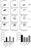
Effect of indoleamine 2,3-dioxygenase (IDO) small interfering (si) RNA on dendritic cell (DC) function, costimulatory and human leucocyte antigen (HLA)-DR expression. (a) Expression of costimulatory and HLA-DR molecules in siRNA-transfected DCs. Immature DCs were transfected with IDO siRNA-3 (1·5 μg/112·5 pmol) or control siRNA (1·5 μg/112·5 pmol) and then they were stimulated with tumour necrosis factor-α (TNF-α) for 2 days to induce maturation. Subsequently, the cells were washed and the expression of the indicated cell surface markers was analysed by flow cytometry using specific monoclonal antibodies. The percentages of positive cells for this representative experiment are shown. (b) siRNA-transfected DCs are capable of processing and presenting soluble antigens to autologous T cells. Irradiated control DCs, control-siRNA-transfected DCs and IDO-siRNA-transfected DCs (5 × 10⁴) were incubated with autologous T cells (105) in the absence or presence of bacillus Calmette–Guérin (BCG) antigens (5 μg/ml) for 5 days. T-cell proliferation was measured by [3H]thymidine incorporation. Samples were assayed in triplicate and the results represent the mean ± SD of two independent experiments. (c) Anti-IDO siRNA inhibits the suppressive effect of IDO-positive DCs on T-cell proliferation. Control DCs, control-siRNA-transfected DCs and IDO-siRNA-transfected DCs were stimulated for 18 hr with IFN-γ to induce IDO expression. Subsequently, they were washed, irradiated and 105 cells were incubated with autologous T cells (104) for 3 days in the presence of phytohaemagglutinin (5 μg/ml). Cell proliferation was measured by [3H]thymidine incorporation. Control 1 = not transfected and IFN-γ-stimulated (IDO-negative DCs), Control 2 = not transfected but IFN-γ-stimulated (IDO-positive DCs), Control siRNA = transfected with control siRNA and IFN-γ-stimulated, IDO siRNA = transfected with IDO siRNA and IFN-γ-stimulated. Samples were assayed in triplicates and the results represent the mean ± SD of two independent experiments.
Figure 6
Figure 6
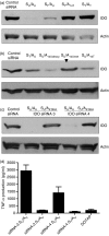
Effect of uridine substitution on small interfering (si) RNA silencing potency. (a) Freshly isolated monocytes were transfected with unmodified and modified indoleamine 2,3-dioxygenase (IDO) siRNA-4 or control siRNA and then stimulated for 18 hr with interferon-γ (IFN-γ; 500 U/ml) to induce IDO expression and IDO protein levels were determined by Western blots. SU/AU = unmodified siRNA, ST/AU = the uridines in the sense strand were replaced by thymidines, ST/A14U = the uridines in the sense strand were replaced by thymidines and only uridine at position 14 in the anti-sense strand was replaced by a thymidine, ST/ST = the uridines in the sense and the anti-sense strands were replaced by thymidines. (b) Effects of site-specific modification at position 14 of the anti-sense strand on siRNA-4 silencing potency. Experimental conditions are as in (a). Modified anti-sense strand carrying either 2′-deoxyuridine, 2′-O-methyl uridine, or thymidine at position 14 was combined with unmodified sense strand to form siRNA duplexes that were tested. As indicated by the arrow, the siRNA with the 2′-O-methyl uridine exhibited no significant silencing effect when compared with the other molecules. (c) Modified IDO siRNAs with a DNA seed sequence were inactive. Anti-IDO siRNA-3 and siRNA-4 with 5′-DNA seed sequences (nucleotides 1–7) were designed and tested in human monocytes. Experimental conditions are as in (a). (d) Effect of thymidines in the sense strand on tumour necrosis factor-α (TNF-α) expression. Unmodified or chemically made siRNAs carrying thymidine only in the sense strand were complexed with the cationic liposome DOTAP (1 μg) and delivered to monocytes. After 18 hr of incubation, TNF-α levels in culture supernatants were analysed by enzyme-linked immunosorbent assay. The final concentration of each siRNA was 1·5 μg (112·5 pmol)/ml. Samples were assayed in triplicates and the results represent the mean ± SD of two independent experiments.
Figure 7
Figure 7
Effects of bifunctional anti-indoleamine 2,3-dioxygenase (IDO) small interfering (si) RNAs on IDO expression, cytokine production, and dendritic cell (DC) function and maturation. (a) Gene silencing using in-vitro-transcribed anti-IDO siRNA-4. Immature monocyte-derived (immo) DCs were transfected with in-vitro-transcribed siRNA-4 (1 or 3 μg/75 or 225 pmol) using the BTX electroporation method and then they were stimulated with interferon-γ (IFN-γ) to induce IDO expression. After 18 hr of transfection, IDO and actin expression were analysed by Western blots. (b and c) Effects of invitro-transcribed siRNA-4 on tumour necrosis factor-α (TNF-α) and interleukin-12 (IL-12) expression. ImmoDC were transfected with in-vitro-transcribed siRNA-4 (3 μg/225 pmol) as in (a). After 18 hr of transfection, TNF-α and IL-12 levels in culture supernatants were analysed by enzyme-linked immunosorbent assay. Samples were assayed in triplicates and the results represent the mean ± SD of two independent experiments. (d) The expression of costimulatory molecules and human leucocyte antigen (HLA) -DR in the transfected immoDCs was determined by flow cytometry after 48 hr of transfection. The percentages of cells for this representative experiment are indicated. (e) The capacity of the 5′-triphosphate siRNA-4-transfected immoDCs to activate T cells was tested using bacillus Calmette–Guérin (BCG) protein lysates. Irradiated DCs (5 × 10⁴) were incubated with autologous T cells (105) in the presence or absence of BCG protein extracts (5 μg/ml) for 5 days. Cell proliferation was measured by [3H]thymidine incorporation. Samples were analysed in triplicates and the results represent the mean ± SD of three independent experiments.

Similar articles

Cited by

References

    1. Banchereau J, Steinman RM. Dendritic cells and the control of immunity. Nature. 1998;392:245–52. - PubMed
    1. Munn DH, Zhou M, Attwood JT, Bondarev I, Conway SJ, Marshall B, Brown C, Mellor AL. Prevention of allogeneic fetal rejection by tryptophan catabolism. Science. 1998;281:1191–3. - PubMed
    1. Munn DH, Mellor AL. IDO expression by dendritic cells: tolerance and tryptophan catabolism. Nat Rev Immunol. 2004;4:762–74. - PubMed
    1. Furset G, Floisand Y, Sioud M. Impaired expression of indoleamine 2, 3-dioxygenase in monocyte-derived dendritic cells in response to Toll-like receptor-7/8 ligands. Immunology. 2008;123:263–71. - PMC - PubMed
    1. Munn DH, Sharma MD, Hou D, et al. Expression of indoleamine 2,3-dioxygenase by plasmacytoid dendritic cells in tumor-draining lymph nodes. J Clin Invest. 2004;114:280–90. - PMC - PubMed

Publication types

MeSH terms

Substances