. 2009 Nov 6;284(45):31085-96.
doi: 10.1074/jbc.M109.035576. Epub 2009 Sep 4.

Mechanism of the internal ribosome entry site-mediated translation of serine hydroxymethyltransferase 1

Affiliations

Mechanism of the internal ribosome entry site-mediated translation of serine hydroxymethyltransferase 1

Jennifer T Fox et al. J Biol Chem. .

Abstract

The 5'-untranslated region (UTR) of serine hydroxymethyltransferase 1 (SHMT1) contains an internal ribosome entry site (IRES) that regulates SHMT1 expression, a rate-limiting enzyme in de novo thymidylate biosynthesis. In this study, we show that the SHMT1 IRES is the first example of a cellular IRES that is poly(A) tail-independent. Interactions between the 5'-UTR and 3'-UTR functionally replaced interactions between the poly(A) tail and the poly(A)-binding protein (PABP) to achieve maximal IRES-mediated translational efficiency. Depletion of the SHMT1 IRES-specific trans-acting factor (ITAF) CUG-binding protein 1 (CUGBP1) from in vitro translation extracts or deletion of the CUGBP1 binding site on the 3'-UTR of the SHMT1 transcript decreased the IRES activity of non-polyadenylylated biscistronic mRNAs relative to polyadenylylated biscistronic mRNAs and resulted in a requirement for PABP. We also identified a novel ITAF, heterogeneous nuclear ribonucleoprotein H2 (hnRNP H2), that stimulates SHMT1 IRES activity by binding to the 5'-UTR of the transcript and interacting with CUGBP1. Collectively, these data support a model for the IRES-mediated translation of SHMT1 whereby the circularization of the mRNA typically provided by the eukaryotic initiation factor (eIF) 4G/PABP/poly(A) tail interaction is achieved instead through the hnRNP H2/CUGBP1-mediated interaction of the 5'- and 3'-UTRs of the SHMT1 transcript. This circularization enhances the IRES activity of SHMT1 by facilitating the recruitment and/or recycling of ribosomal subunits, which bind to the transcript in the middle of the 5'-UTR and migrate to the initiation codon via eIF4A-mediated scanning.


Figures

FIGURE 1.
FIGURE 1.
The poly(A) tail and PABP are not required for maximal SHMT1 IRES activity. A, the bicistronic mRNA used to quantify SHMT1 IRES activity. It consists of (in the 5′ to 3′ direction) a cap analog, the Rluc reporter gene, the alternatively spliced form of the human SHMT1 5′-UTR lacking exon 2 (25), the Fluc reporter gene, and where indicated, the full-length human SHMT1 3′-UTR and a 30-nucleotide poly(A) tail (25). B, in vitro translation assays were carried out using rabbit reticulocyte lysate and in vitro transcribed bicistronic mRNAs with (5′-UTR + 3′-UTR) and without (5′-UTR) the SHMT1 3′-UTR. The white bars represent the ratio of IRES-mediated translation (Fluc) to cap-dependent translation (Rluc) of bicistronic mRNA containing a 30-nucleotide poly(A) tail, and the dark bars represent the Fluc/Rluc of bicistronic mRNA lacking a poly(A) tail. The relative ratio for each bicistronic mRNA containing a poly(A) tail was given a value of 1.0. The data represent the average of three independent experiments ± S.E. C, the bicistronic mRNAs described in A were labeled with 32P, and in vitro translation assays were performed as described under ”Experimental Procedures.“ The RNAs were resolved on an agarose gel and transferred to a positively charged nylon membrane. For each transcript, the left lane represents the mRNA before the in vitro translation reaction, and the right lane represents the transcript after the in vitro translation reaction. D, rabbit reticulocyte lysate was incubated with either GST (Control) or GST-Paip2 (PABP-depleted). The depletion of PABP by GST-Paip2 but not GST alone was confirmed by immunoblotting (right) using an antibody against PABP. GAPDH served as a control for equal protein loading. The graph on the left shows the relative IRES activity (as measured by Fluc/Rluc) of the bicistronic mRNAs in control (striped bars) and PABP-depleted (black bars) rabbit reticulocyte lysate. The relative luminosity for each bicistronic mRNA in the control reaction was given a value of 1.0. The data represent the average of three independent experiments ± S.E.
FIGURE 2.
FIGURE 2.
Proposed model for the IRES-mediated translation of SHMT1. In this model, an interaction between an ITAF bound to the 5′-UTR of the SHMT1 transcript and another ITAF bound to the 3′-UTR of the SHMT1 transcript serves to circularize the mRNA. This results in the formation of a closed loop similar to the one that is typically formed by the eIF4G/PABP/poly(A) tail interaction.
FIGURE 3.
FIGURE 3.
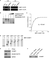
The interaction of CUGBP1 with the SHMT1 UTRs. A, RNA was immunoprecipitated from UV-treated MCF-7 whole cell extract using an antibody against IgG (control for nonspecific binding) or CUGBP1. The RNA in lanes 1–3 (+RT) was reverse-transcribed into cDNA and then analyzed by PCR using primers specific to either the SHMT1 5′-UTR or the SHMT1 3′-UTR. The RNA in lanes 4–6 (−RT) did not undergo the reverse transcription step. Rather, they were analyzed directly by PCR to control for DNA contamination in the immunoprecipitates. Lanes 1 and 4 (Input) represent 1% of the RNA used in the immunoprecipitation. B, electrophoretic mobility shift assays were carried out in the presence of excess yeast tRNA to eliminate nonspecific binding of the probe, radiolabeled SHMT1 3′-UTR, and recombinant MBP-CUGBP1. A 10×, 50×, and 100× molar excess of unlabeled SHMT1 3′-UTR was also added in lanes 3, 4, and 5, respectively, to determine binding specificity. C, electrophoretic mobility shift assays were carried out using radiolabeled SHMT1 3′-UTR and increasing concentrations of MBP-CUGBP1. The fraction of the 3′-UTR bound by the recombinant protein was quantified using ChemiImager 4400 from Alpha Innotech Corp. The dissociation constant (Kd) was determined using GraphPad Prism. D, electrophoretic mobility shift assays were carried out using radiolabeled SHMT1 3′-UTR truncation mutants in the absence and presence of MBP-CUGBP1. The nucleotides of the 3′-UTR that comprise the truncation mutant are listed above each gel. The nucleotide at the 5′-end of the 3′-UTR is labeled 1. The nucleotide at the 3′-end of the 3′-UTR is labeled 637. E, MCF-7 whole cell extract was either not treated or treated with λ-phosphatase (PPase) and analyzed by Western blot using antibodies against CUGBP1 and phosphorylated serine and threonine residues. GAPDH served as a control for equal protein loading. F, the extracts from E were applied to RNA affinity columns to which either the SHMT1 5′-UTR or the RevUTR had been attached. Proteins that bound to the UTRs were eluted and analyzed by Western blotting using an antibody against CUGBP1.
FIGURE 4.
FIGURE 4.
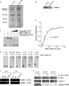
Interaction of hnRNP H2 with the SHMT1 UTRs. A, MCF-7 whole cell extract was applied to RNA affinity columns to which either the SHMT1 5′-UTR or the RevUTR had been attached. Proteins that bound to the UTRs were eluted, separated by SDS-PAGE, and visualized by Coomassie Blue staining. The protein marked with an asterisk was excised from the gel and analyzed by μLC/MS/MS. B, the SDS-polyacrylamide gel from A was transferred to a PVDF membrane and analyzed by Western blotting using an antibody against hnRNP H. C, electrophoretic mobility shift assays were carried out in the presence of excess yeast tRNA to eliminate nonspecific binding of the probe, radiolabeled SHMT1 5′-UTR, and recombinant MBP-hnRNP H2. A 10×, 50×, and 100× molar excess of unlabeled SHMT1 5′-UTR was also added in lanes 3, 4, and 5, respectively, to determine binding specificity. D, electrophoretic mobility shift assays were carried out using radiolabeled SHMT1 5′-UTR and increasing concentrations of MBP-hnRNP H2. The fraction of the 5′-UTR bound by the recombinant protein was quantified using ChemiImager 4400 from Alpha Innotech Corp. The dissociation constant (Kd) was determined using GraphPad Prism. E, electrophoretic mobility shift assays were carried out using radiolabeled SHMT1 5′-UTR truncation mutants in the absence and presence of MBP-hnRNP H2. The nucleotides of the 5′-UTR that comprise the truncation mutant are listed above each gel. The nucleotide at the 5′-end of the 5′-UTR is labeled 1. The nucleotide at the 3′-end of the 5′-UTR is labeled 190. F, RNA was immunoprecipitated from UV-treated MCF-7 whole cell extract using an antibody against IgG (control for nonspecific binding) or hnRNP H. The RNA in lanes 1–3 (+RT) was reverse-transcribed into cDNA and then analyzed by PCR using primers specific to either the SHMT1 5′-UTR or the SHMT1 3′-UTR. The RNA in lanes 4–6 (−RT) did not undergo the reverse transcription step. Rather, they were analyzed directly by PCR to control for DNA contamination in the immunoprecipitates. Lanes 1 and 4 (Input) represent 1% of the RNA used in the immunoprecipitation. G, whole cell extract (WCE) from cells that were treated with either negative control siRNA (−CUGBP1 siRNA) or siRNA directed against CUGBP1 were applied to RNA affinity columns to which either the cSHMT 5′-UTR or the cSHMT 3′-UTR had been attached. Proteins that bound to the UTRs were eluted, separated by SDS-PAGE, and visualized by Western blotting using antibodies against CUGBP1 or hnRNP H. For the whole cell extract, equal protein loading was confirmed by using an antibody against GAPDH. For the affinity column elutions, equal protein loading was confirmed by staining the gel with Coomassie blue after transfer.
FIGURE 5.
FIGURE 5.
hnRNP H2 binds to CUGBP1 in an RNA-independent manner. CUGBP1 (A) and hnRNP H (B) were coimmunoprecipitated from MCF-7 whole cell extracts using antibodies against hnRNP H and CUGBP1, respectively. The no antibody, IgG, and hemagglutinin (HA) coimmunoprecipitations serve as controls for nonspecific binding. CUGBP1 (C) and hnRNP H (D) were coimmunoprecipitated from a mixture of recombinant hnRNP H2 and recombinant CUGBP1 using antibodies against hnRNP H and CUGBP1, respectively. The no antibody and IgG coimmunoprecipitations serve as controls for nonspecific binding.
FIGURE 6.
FIGURE 6.
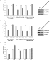
CUGBP1 and hnRNP H2 depletion result in a dependence on the poly(A) tail. CUGBP1 (A) and hnRNP H (B) were immunodepleted from rabbit reticulocyte lysate as described under ”Experimental Procedures.“ The depletion of these proteins was confirmed by immunoblotting (right) using antibodies against CUGBP1 (A) and hnRNP H (B). GAPDH served as a control for equal protein loading. The graphs on the left show the IRES activities of the bicistronic mRNAs containing a 30-nucleotide poly(A) tail (white bars) or lacking a poly(A) tail (dark bars) as measured in control rabbit reticulocyte, CUBBP1 (A)- or hnRNP H (B)-depleted rabbit reticulocyte lysate, or immunodepleted lysate supplemented with recombinant CUGBP1 (A) or hnRNP H2 (B). The relative ratio of Fluc/Rluc for each bicistronic mRNA containing a poly(A) tail was given a value of 1.0. The data represent the average of three independent experiments ± S.E. C, the 3′-UTR of the bicistronic mRNA was truncated by removal of nucleotides from the 3′-end, and the IRES activity of these truncation mutants was measured in rabbit reticulocyte lysate. The white bars represent the IRES activity of the truncated bicistronic mRNA containing a 300nucleotide poly(A) tail, and the shaded bars represent the IRES activity of the truncated bicistronic mRNA lacking a poly(A) tail. The relative ratio for each truncated bicistronic mRNA containing a poly(A) tail was given a value of 1.0. The data represent the average of three independent experiments ± S.E.
FIGURE 7.
FIGURE 7.
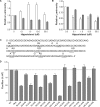
Ribosome scanning occurs between nucleotides 103 and 118 of the SHMT1 5′-UTR. MCF-7 cells were transiently transfected with polyadenylylated bicistronic mRNAs containing the hepatitis C virus IRES (A) or the SHMT1 5′-UTR (B) and then treated with the indicated amount of hippuristanol. Following 11 h of treatment, Fluc (white bars) and Rluc (dark bars) expression was quantified as described under ”Experimental Procedures.“ The relative luminosity in untreated cells was given a value of 1.0. The data represent the average of three independent experiments ± S.E. C, the sequence of the SHMT1 5′-UTR indicating the positions of the inserted open reading frames. The location of each start and stop codon is underlined, and the letters above the underlined nucleotides indicate changes to the wild-type sequence that were made by site-directed mutagenesis. D, MCF-7 cells were transiently transfected with bicistronic mRNAs containing the mutated SHMT1 5′-UTRs. The number of each mutant represents the position of the A in the AUG or AUA, or the U in the UUG. The relative ratio of Fluc/Rluc for each the wild-type (WT) bicistronic mRNA was given a value of 100%. The data represent the average of at least three independent experiments ± S.E.

Similar articles

Cited by

References

    1. Jang S. K., Kräusslich H. G., Nicklin M. J., Duke G. M., Palmenberg A. C., Wimmer E. (1988) J. Virol. 62, 2636–2643 - PMC - PubMed
    1. Pelletier J., Sonenberg N. (1988) Nature 334, 320–325 - PubMed
    1. Hellen C. U., Sarnow P. (2001) Genes Dev. 15, 1593–1612 - PubMed
    1. Stoneley M., Willis A. E. (2004) Oncogene 23, 3200–3207 - PubMed
    1. Johannes G., Carter M. S., Eisen M. B., Brown P. O., Sarnow P. (1999) Proc. Natl. Acad. Sci. U.S.A. 96, 13118–13123 - PMC - PubMed

Publication types