. 2009 Sep 8;16(3):232-45.
doi: 10.1016/j.ccr.2009.07.030.

The B cell mutator AID promotes B lymphoid blast crisis and drug resistance in chronic myeloid leukemia

Affiliations

The B cell mutator AID promotes B lymphoid blast crisis and drug resistance in chronic myeloid leukemia

Lars Klemm et al. Cancer Cell. .

Abstract

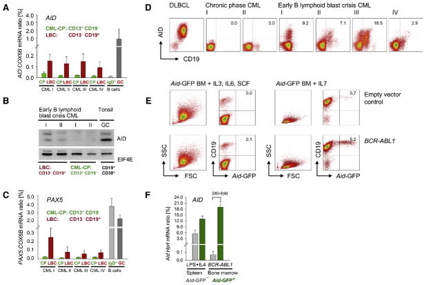
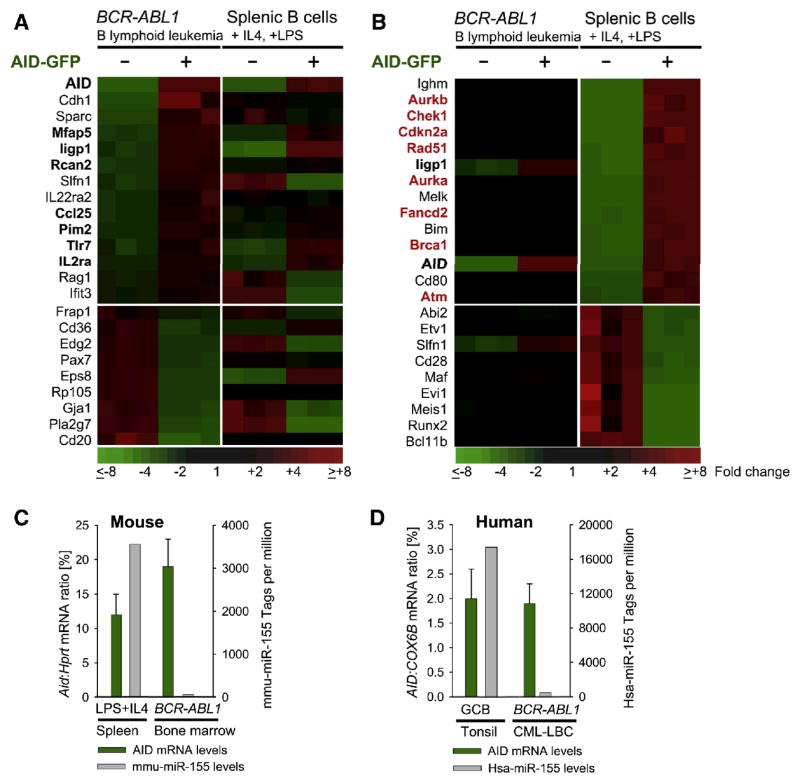
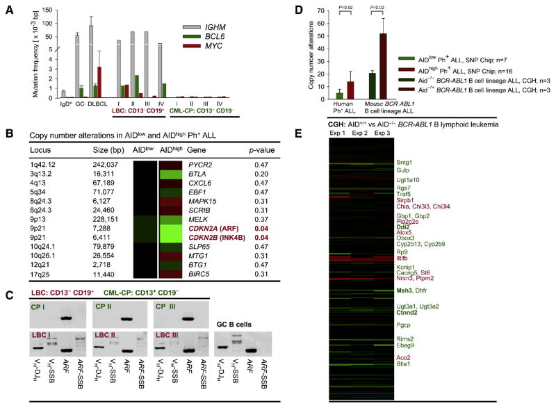
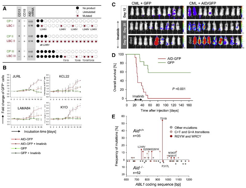
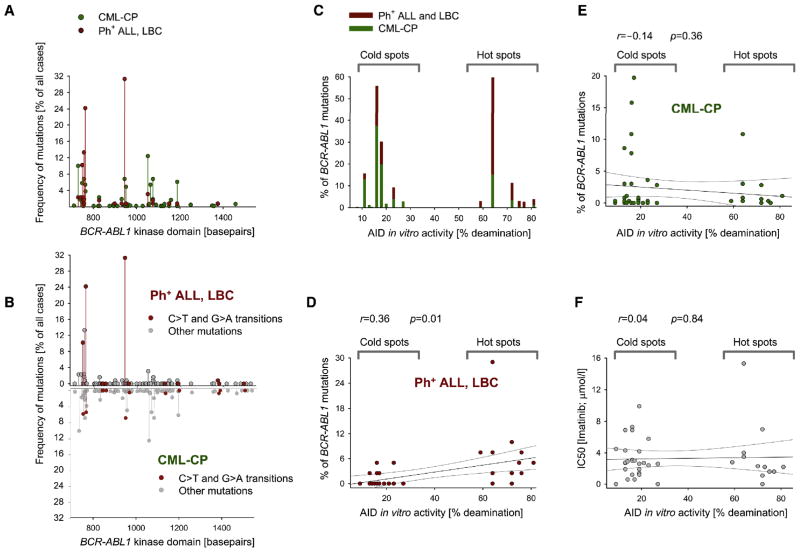
Chronic myeloid leukemia (CML) is induced by BCR-ABL1 and can be effectively treated for many years with Imatinib until leukemia cells acquire drug resistance through BCR-ABL1 mutations and progress into fatal B lymphoid blast crisis (LBC). Despite its clinical significance, the mechanism of progression into LBC is unknown. Here, we show that LBC but not CML cells express the B cell-specific mutator enzyme AID. We demonstrate that AID expression in CML cells promotes overall genetic instability by hypermutation of tumor suppressor and DNA repair genes. Importantly, our data uncover a causative role of AID activity in the acquisition of BCR-ABL1 mutations leading to Imatinib resistance, thus providing a rationale for the rapid development of drug resistance and blast crisis progression.


Conflict of interest statement

The authors have no conflicting financial interests.

Figures

Figure 1
Figure 1. B cell lineage-specific activation of AID in BCR-ABL1-transformed leukemia cells
(A) Quantitative RT-PCR analysis of AID mRNA expression in CD13 CD19+ B lymphoid (CML-LBC) and CD13+ CD19 myeloid (CML-CP) bone marrow cells sorted from four patients (I-IV) with early B lymphoid blast crisis CML. Peripheral blood naïve (IgD+) and germinal center (GC) B cells were used as negative and positive controls, respectively. AID mRNA levels (means ± SD) were normalized based on COX6B transcripts. (B) AID protein levels in CML-LBC and CML-CP cells from two patients (I-II) were compared to those of tonsillar GC B cells by Western blotting using EIF4E as loading control. (C) PAX5 mRNA levels in sorted CML-CP and CML-LBC cells were measured by quantitative RT-PCR (means + SD). (D) AID protein expression in chronic phase and lymphoid blast crisis CML cells (I-IV) was measured by flow cytometry (intracellular staining) using diffuse large B cell lymphoma (DLBCL) cells as control. (E) Bone marrow cells from AID-GFP reporter transgenic mice were transduced with BCR-ABL1 or empty vector control and cultured under myeloid (IL3, IL6, SCF) or B lymphoid (IL7) growth conditions. (F) AID mRNA levels (means ± SD) in sorted GFP (gray) and GFP+ (green) cell populations from LPS/IL4-stimulated splenic B cells or BCR-ABL1-transformed leukemia cells from AID-GFP transgenic mice were measured by quantitative RT-PCR
Figure 2
Figure 2. BCR-ABL1-transformed B lymphoid leukemia cells express AID in the absence of protective mechanisms to maintain genome integrity
Bone marrow cells and splenic B cells were isolated from Aid-GFP reporter transgenic mice. Bone marrow B cell precursors were transformed with retroviral BCR-ABL1, splenic B cells were activated by LPS and IL4. From BCR-ABL1-transformed leukemia cells and LPS/IL4-activated B cells, Aid-GFP+ and Aid-GFP cells were sorted and subjected to RNA isolation and microarray analysis using the 430 GeneChip platform (GEO accession number GSE13611). For both cell types, genes that were differentially expressed between Aid-GFP+ and Aid-GFP populations were identified and sorted according to the ratio of gene expression values in Aid-GFP+ and Aid-GFP- populations from BCR-ABL1-transformed leukemia cells (A) and LPS/IL4-activated B cells (B). Genes that are concordantly upregulated with AID expression both in BCR-ABL1 leukemia (A) and activated splenic B cells (B) are highlighted in boldface. Genes related to DNA damage and repair and chromosomal stability are highlighted in red. In (C) and (D), AID mRNA levels (% of reference gene (means ± SD) as determined by quantitative RT-PCR) were plotted against expression levels of miR-155 (microRNA tags per million, as determined by Illumina deep sequencing). To this end, Aid mRNA and mmu-miR-155 levels were measured in murine LPS/IL4-activated splenic B cells and mouse bone marrow B cell precursors that were transformed by BCR-ABL1 (C). Likewise, AID mRNA and hsa-miR-155 levels were measured in sorted human tonsillar germinal center B cells (GCB; CD77highCD38+IgD) and human B lymphoid CML-LBC cells (D).
Figure 3
Figure 3. Evidence of aberrant AID activity in B lymphoid BCR-ABL1 leukemias
(A) CML-LBC (CD13 CD19+ CD34+) and CML-CP (CD13+CD19 CD34+) cells were sorted from four patients and analyzed for somatic mutations within rearranged IGHM loci, as well as BCL6 and MYC genes (means of mutation frequencies ± SD). Mutation frequencies in naïve (IgD+) and germinal center (GC) B cells, as well as DLBCL cells were studied as a reference. CML-CP cells carry IGHM loci in germline configuration, which precludes mutation analysis. (B) SNP mapping analysis was performed for 23 bone marrow samples from patients with B lymphoid Ph+ ALL (GEO accession number GSE13612). Samples were selected based on particularly high (n=16; AIDhigh) or low (n=7; AIDlow) AID mRNA levels. Deletions or duplications of gene loci are represented in green or red respectively, with color intensities reflecting the frequency of these lesions based on the total number of samples analyzed. (C) Short-lived DNA single-strand break (SSB) intermediates were determined at the ARF and IGHM loci by ligation-mediated PCR (LM-PCR) in LBC and CML-CP, as well as in germinal center B cells (GC). The presence of ARF and rearranged VH-DJH genes was verified by genomic PCR in each sample. (D) Gene copy number alterations (CNA) were studied in BCR-ABL1-transformed bone marrow B cell precursors from AID−/− and AID+/+ mice by Comparative Genomic Hybridization (means of CNA frequencies + SD; GEO accession number GSE15093) using genomic DNA from normal splenic B cells as a reference. AID-specific lesions were identified in three CGH experiments comparing AID+/+ BCR-ABL1 leukemia cells against AID−/− BCR-ABL1 leukemia (E). Data for all genes/loci where a copy number change between AID−/− and AID+/+ BCR-ABL1 leukemia was found in at least one experiment (sorted by chromosomal localization, chromosomes 1 through 19; recurrent deletions (green) or amplifications (red) are highlighted; E).
Figure 4
Figure 4. AID expression in CML cells induces Imatinib-resistance in vitro and in vivo
(A) Single CML-CP and LBC cells from three patients (I-III) with early lymphoid blast crisis under Imatinib-treatment were sorted into individual PCR reaction tubes and studied by single-cell PCR. AID mRNA levels (percentage of COX6B mRNA) for each cell population is indicated. (B) Four AID-negative human myeloid CML cell lines were transduced with retroviral vectors encoding AID-GFP or GFP alone and cultured for three weeks in the presence or absence of 2 μmol/l Imatinib. Relative outgrowth or depletion of GFP+ cells was monitored by flow cytometry (means ± SD). (C) Firefly luciferase-labeled AID-GFP+ and GFP+ CML cells were intrafemorally injected into sublethally irradiated NOD/SCID recipient mice. Leukemia cell growth was monitored by bioluminescence imaging at the times indicated. (E) Overall survival of NOD/SCID mice injected with either AID-GFP+ or GFP+ CML cells and treated with 100 mg/kg/day Imatinib (day 20–40) is depicted by a Kaplan-Meier analysis. (F) Bone marrow B cell precursor cells from AID+/+ and AID−/− mice were transformed by BCR-ABL1 and cultured for 6 weeks under B lymphoid growth conditions. Subsequently, AID+/+ and AID−/− BCR-ABL1 B lymphoid leukemia cells were treated with gradually increasing concentrations of Imatinib (0.1 to 1.75 μmol/l) for two weeks. Surviving cells were subjected to sequence analysis for BCR -ABL1 kinase mutations. Red circles: ABL1 mutations conferring clinical Imatinib-resistance, orange circles: C→T or G→A transitions, gray circles: other mutations.
Figure 5
Figure 5. Ectopic expression of PAX5 in CML cells induces partial B cell lineage conversion, AID expression and Imatinib-resistance
(A) CML cell lines were transduced with retroviral vectors encoding GFP (green) or PAX5/GFP (red) and expression of B cell lineage specific genes PAX5, AID, CD79A (Igα) and SLP65 (BLNK) was monitored by quantitative RT-PCR (means ± SD). (B) FACS analysis of CD19+ subclones emerging from GFP or PAX5/GFP-transduced CML cells cultured in the presence or absence of Imatinib. (C). Spectratyping analysis of rearranged immunoglobulin heavy chain genes from GFP-transduced (left), PAX5/GFP-transduced CML cells (middle), or primary CD19+ peripheral blood B cells pooled from four healthy donors (right). The bottom panel shows the sequence analysis of oligoclonal IGHM gene rearrangements amplified from PAX5/GFP-transduced CML cells. (D) Four different CML cell lines were transduced with GFP or PAX5/GFP and grown in the presence or absence of rising concentrations of Imatinib (from 0.1 μmol/l to 1.75 μmol/l) for 16 days. The percentage of GFP + cells (means ± SD) was monitored over time by flow cytometry.
Figure 6
Figure 6. Characteristics of BCR-ABL1 kinase domain mutations in myeloid chronic phase CML and B lymphoid Ph+ALL/CML lymphoid blast crisis
In (A), the distribution of BCR-ABL1 kinase mutations in CML-CP (n=572; green) as compared to Ph+ ALL/LBC (n=128; red) is compared. The x-axis denotes the nucleotide position of mutations. In (B), the frequencies of BCR-ABL1 mutations in B lymphoid Ph+ALL/LBC (top) and myeloid CML-CP (bottom) are compared with respect to C→T and G→A transitions (red). As a quantitative measure of AID-specific mutability, data from a comprehensive AID in vitro deamination assay based on 68 C-based motifs (Yu et al., 2004) were used. Motifs with AID in vitro activity below 27% and above 59% were considered coldspots and hotspots, respectively. In (C–E), the distribution of BCR-ABL1 kinase mutations in B lymphoid Ph+ALL/LBC (red) and myeloid CML-CP (green) are compared with respect to AID in vitro activity at the site of mutation (% deamination; based on data from Yu et al., 2004). In (C), the frequency of mutations at AID hotspot and AID coldspot motifs are compared in Ph+ALL/LBC (red) and myeloid CML-CP (green). A potential correlation between the frequency of BCR-ABL1 mutations and AID-specific mutability at these sites (% deamination) was tested separately for Ph+ALL/LBC (red; D) and CML-CP (green; E). In (F) the possibility of a potential bias introduced into our analysis is tested: Mutations with a high IC50 for Imatinib confer a high degree of drug-resistance and, hence, a strong selective survival advantage, which may skew the pattern of mutations (e.g. frequency of AID hotspot targeting). For 41 unique mutations, information on both IC50 (Imatinib) as well as AID in vitro activity (% deamination; Yu et al., 2004) was available (F). Correlation coefficients (r) and p-values were calculated based on Pearson product moment correlation. Linear regressions and 95% confidence intervals are indicated by black and gray lines, respectively.

Comment in

Similar articles

Cited by

References

    1. Blagodatski A, Batrak V, Schmidl S, Schoetz U, Caldwell RB, Arakawa H, Buerstedde JM. A cis-acting diversification activator both necessary and sufficient for AID-mediated hypermutation. PLoS Genet. 2009;5:e1000332. - PMC - PubMed
    1. Calabretta B, Perrotti D. The biology of CML blast crisis. Blood. 2004;103:4010–22. - PubMed
    1. Casellas R, Yamane A, Kovalchuk AL, Potter M. Restricting activation-induced cytidine deaminase tumorigenic activity in B lymphocytes. Immunology. 2009;126:316–28. - PMC - PubMed
    1. Chaudhuri J, Tian M, Khuong C, Chua K, Pinaud E, Alt FW. Transcription-targeted DNA deamination by the AID antibody diversification enzyme. Nature. 2003;422:726–730. - PubMed
    1. Chesi M, Robbiani DF, Sebag M, Chang WJ, Affer M, Tiedemann R, Valdez R, Palmer SE, Haas SS, Stewart AK, Fonseca R, et al. AID-dependent activation of a MYC transgene induces multiple myeloma in a conditional mouse model of post-germinal center malignancies. Cancer Cell. 2008;13:167–80. - PMC - PubMed

Publication types

MeSH terms

Associated data