

Alveolar macrophage dysregulation in Hermansky-Pudlak syndrome type 1
- PMID: 19729668
- PMCID: PMC2784416
- DOI: 10.1164/rccm.200901-0023OC
Alveolar macrophage dysregulation in Hermansky-Pudlak syndrome type 1
Abstract
Rationale: Individuals with Hermansky-Pudlak syndrome type 1 (HPS-1), an autosomal recessive disorder characterized by defective biogenesis of lysosome-related organelles, develop an accelerated form of progressive fibrotic lung disease. The etiology of pulmonary fibrosis associated with HPS-1 is unknown.
Objectives: To investigate the potential pathogenesis of pulmonary fibrosis in HPS-1, lung cells and proteins from individuals with HPS-1 were studied.
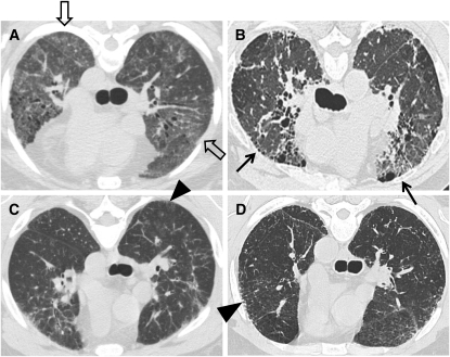
Methods: Forty-one subjects with HPS-1 with and without pulmonary fibrosis were evaluated with pulmonary function tests, high-resolution computed tomography scan, and bronchoscopy. Bronchoalveolar lavage cells and analytes were analyzed.
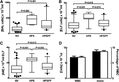
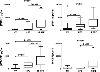
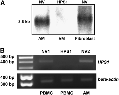
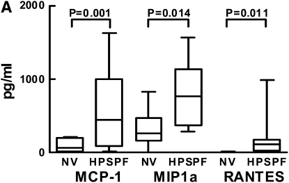
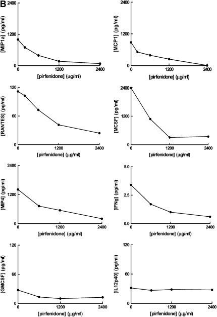
Measurements and main results: Concentrations of total bronchoalveolar lavage cells and alveolar macrophages were significantly higher in epithelial lining fluid from subjects with HPS-1 with and without pulmonary fibrosis compared with healthy research volunteers. Concentrations of cytokines and chemokines (i.e., monocyte chemoattractant protein-1, macrophage inflammatory protein-1alpha, and granulocyte-macrophage colony-stimulating factor) in alveolar epithelial lining fluid were significantly higher in subjects with HPS-1 with and without pulmonary fibrosis compared with healthy research volunteers (P < 0.001). In vitro, HPS-1 pulmonary fibrosis alveolar macrophages, which did not express HPS1 mRNA, secreted significantly higher concentrations of monocyte chemoattractant protein-1, macrophage inflammatory protein-1alpha, and regulated upon activation, normal T cell expressed and secreted (RANTES) protein compared with normal cells (P = 0.001, P = 0.014, and P = 0.011, respectively). Pirfenidone suppressed HPS-1 alveolar macrophage cytokine and chemokine secretion in vitro in a dose-dependent manner.
Conclusions: In HPS-1, alveolar inflammation predominantly involves macrophages and is associated with high lung concentrations of cytokines and chemokines. HPS-1 alveolar macrophages provide a model system in which to study the pathogenesis and treatment of HPS pulmonary fibrosis.
Figures
Similar articles
-
The alveolar epithelium determines susceptibility to lung fibrosis in Hermansky-Pudlak syndrome.Am J Respir Crit Care Med. 2012 Nov 15;186(10):1014-24. doi: 10.1164/rccm.201207-1206OC. Epub 2012 Oct 4. Am J Respir Crit Care Med. 2012. PMID: 23043085 Free PMC article.
-
Early alveolar epithelial dysfunction promotes lung inflammation in a mouse model of Hermansky-Pudlak syndrome.Am J Respir Crit Care Med. 2011 Aug 15;184(4):449-58. doi: 10.1164/rccm.201011-1882OC. Am J Respir Crit Care Med. 2011. PMID: 21616998 Free PMC article.
-
Dysregulation of galectin-3. Implications for Hermansky-Pudlak syndrome pulmonary fibrosis.Am J Respir Cell Mol Biol. 2014 Mar;50(3):605-13. doi: 10.1165/rcmb.2013-0025OC. Am J Respir Cell Mol Biol. 2014. PMID: 24134621 Free PMC article.
-
Pathogenesis and Therapy of Hermansky-Pudlak Syndrome (HPS)-Associated Pulmonary Fibrosis.Int J Mol Sci. 2024 Oct 19;25(20):11270. doi: 10.3390/ijms252011270. Int J Mol Sci. 2024. PMID: 39457053 Free PMC article. Review.
-
Hermansky-Pudlak Syndrome.Clin Chest Med. 2016 Sep;37(3):505-11. doi: 10.1016/j.ccm.2016.04.012. Epub 2016 Jun 30. Clin Chest Med. 2016. PMID: 27514596 Free PMC article. Review.
Cited by
-
Rare interstitial lung diseases: a narrative review.J Thorac Dis. 2024 Sep 30;16(9):6320-6338. doi: 10.21037/jtd-24-450. Epub 2024 Sep 26. J Thorac Dis. 2024. PMID: 39444900 Free PMC article. Review.
-
Hermansky-Pudlak syndrome interstitial pneumonia: it's the epithelium, stupid!Am J Respir Crit Care Med. 2012 Nov 15;186(10):939-40. doi: 10.1164/rccm.201210-1771ED. Am J Respir Crit Care Med. 2012. PMID: 23155210 Free PMC article. No abstract available.
-
Cellular and Molecular Control of Lipid Metabolism in Idiopathic Pulmonary Fibrosis: Clinical Application of the Lysophosphatidic Acid Pathway.Cells. 2023 Feb 8;12(4):548. doi: 10.3390/cells12040548. Cells. 2023. PMID: 36831215 Free PMC article. Review.
-
Bronchoalveolar lavage as a diagnostic procedure: a review of known cellular and molecular findings in various lung diseases.J Thorac Dis. 2020 Sep;12(9):4991-5019. doi: 10.21037/jtd-20-651. J Thorac Dis. 2020. PMID: 33145073 Free PMC article. Review.
-
Genetic Regulation of Cytokine Response in Patients with Acute Community-Acquired Pneumonia.Genes (Basel). 2022 Jan 6;13(1):111. doi: 10.3390/genes13010111. Genes (Basel). 2022. PMID: 35052452 Free PMC article.
References
-
- Gahl WA, Brantly M, Kaiser-Kupfer MI, Iwata F, Hazelwood S, Shotelersuk V, Duffy LF, Kuehl EM, Troendle J, Bernardini I. Genetic defects and clinical characteristics of patients with a form of oculocutaneous albinism (Hermansky-Pudlak syndrome). N Engl J Med 1998;338:1258–1264. - PubMed
-
- Brantly M, Avila NA, Shotelersuk V, Lucero C, Huizing M, Gahl WA. Pulmonary function and high-resolution CT findings in patients with an inherited form of pulmonary fibrosis, Hermansky-Pudlak syndrome, due to mutations in HPS-1. Chest 2000;117:129–136. - PubMed
-
- Gahl WA, Brantly M, Troendle J, Avila NA, Padua A, Montalvo C, Cardona H, Calis KA, Gochuico B. Effect of pirfenidone on the pulmonary fibrosis of Hermansky-Pudlak syndrome. Mol Genet Metab 2002;76:234–242. - PubMed
-
- Lederer DJ, Kawut SM, Sonett JR, Vakiani E, Seward SL Jr, White JG, Wilt JS, Marboe CC, Gahl WA, Arcasoy SM. Successful bilateral lung transplantation for pulmonary fibrosis associated with the Hermansky-Pudlak syndrome. J Heart Lung Transplant 2005;24:1697–1699. - PubMed
Publication types
MeSH terms
Substances
Grants and funding
LinkOut - more resources
Full Text Sources
Research Materials
