. 2009 Sep;119(9):2772-86.
doi: 10.1172/JCI36154. Epub 2009 Aug 10.

MicroRNA-208a is a regulator of cardiac hypertrophy and conduction in mice

Affiliations

MicroRNA-208a is a regulator of cardiac hypertrophy and conduction in mice

Thomas E Callis et al. J Clin Invest. 2009 Sep.

Abstract

MicroRNAs (miRNAs) are a class of small noncoding RNAs that have gained status as important regulators of gene expression. Here, we investigated the function and molecular mechanisms of the miR-208 family of miRNAs in adult mouse heart physiology. We found that miR-208a, which is encoded within an intron of alpha-cardiac muscle myosin heavy chain gene (Myh6), was actually a member of a miRNA family that also included miR-208b, which was determined to be encoded within an intron of beta-cardiac muscle myosin heavy chain gene (Myh7). These miRNAs were differentially expressed in the mouse heart, paralleling the expression of their host genes. Transgenic overexpression of miR-208a in the heart was sufficient to induce hypertrophic growth in mice, which resulted in pronounced repression of the miR-208 regulatory targets thyroid hormone-associated protein 1 and myostatin, 2 negative regulators of muscle growth and hypertrophy. Studies of the miR-208a Tg mice indicated that miR-208a expression was sufficient to induce arrhythmias. Furthermore, analysis of mice lacking miR-208a indicated that miR-208a was required for proper cardiac conduction and expression of the cardiac transcription factors homeodomain-only protein and GATA4 and the gap junction protein connexin 40. Together, our studies uncover what we believe are novel miRNA-dependent mechanisms that modulate cardiac hypertrophy and electrical conduction.


Figures

Figure 1
Figure 1. Expression of miR-208a and miR-208b parallels the expression of their respective host genes Myh6 and Myh7.
(A) miR-208a is encoded by intron 29 of the Myh6 gene, while miR-208b is encoded by intron 31 of the Myh7 gene. miR-208a and miR-208b are highly conserved and share similar sequence identity (indicated by asterisks). (B) Detection of mature and precursor miR-208a in adult mouse tissues. Sk. muscle, skeletal muscle; tRNA, transfer RNA. (C) Detection of mature and precursor miR-208a in E13.5, E16.5, and neonatal tissues using Northern blot analysis. (D) Top left: αMHC and βMHC transcripts were detected in E16.5, P0, P5, P10, and adult mouse hearts using RT-PCR. Bottom left: miR-208a and miR-208b expression was detected in the samples using Northern analysis. Right: Relative levels of miR-208a and miR-208b during heart development (E) Top left: αMHC and βMHC transcripts were detected using RT-PCR in isolated rat neonatal cardiomyocytes following treatment with thyroid hormone (T3). Bottom left: miR-208a and miR-208b were detected using Northern analysis. Right: Quantitative analysis. Fold change is relative to no T3 treatment (which was set at 1). *P < 0.01, compared with no T3 treatment.
Figure 2
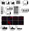
Figure 2. Hearts of miR-208a Tg mice undergo hypertrophic growth.
(A) Left: Northern blot analysis showing an approximately 4-fold increase of miR-208a expression in hearts of miR-208a Tg animals compared with control littermates. U6 served as loading control. Right: Quantitative analysis of fold change in miR-208a expression compared with control animals. *P < 0.001. (B) Gross morphology of miR-208a Tg hearts was enlarged compared with control hearts. Scale bar: 1 mm. (C) Heart weight to body weight ratios (Hw/Bw) of 4-month-old miR-208a Tg mice (n = 22) were significantly higher than controls (n = 19) (P < 7 × 10⁻⁷). (D) Macroscopic view of H&E-stained histological sections (upper, sagittal; lower, transverse) from control and miR-208a Tg hearts. Scale bars: 1 mm. (E) Sarcomeric structure of cardiomyocytes visualized by desmin staining of histological sections. Original magnification, ×200. (F) Histological sections were stained with wheat germ agglutinin–TRITC conjugate to determine cell size. Mean cardiomyocyte size of miR-208a Tg hearts (n = 930) was significantly larger than control cardiomyocytes (n = 926) (*P < 9 × 10–50). Original magnification, ×200. (G) Distribution of control and miR-208a Tg cardiomyocyte cell area measurements were compared. (H) Representative M-mode echocardiographs from conscious control and miR-208a Tg mice. IVSTD, interventricular septal thickness in diastole; IVSTS, interventricular septal thickness in systole; PWTD, posterior wall thickness in diastole; PWTS, posterior wall thickness in systole; LVEDD, left ventricular end-diastolic dimension; LVESD, left ventricular end-systolic dimension.
Figure 3
Figure 3. miR-208a overexpression induces hypertrophic gene expression.
(A) Transcripts for αMHC, βMHC, and ANF were detected by real-time PCR in 4-month-old hearts from control and miR-208a Tg mice (n = 5 per genotype). Data are the mean fold change in expression ± SEM. *P < 0.01. (B) Left: Western blot analysis of total MHC and βMHC protein levels in adult control and miR-208a Tg hearts. Right: Quantitative analysis of fold change in protein levels. *P < 0.01. (C) Top left: RT-PCR was used to detect αMHC, βMHC, and ANF transcripts in wild-type hearts following 3 weeks thoracic aortic banding (TAB) or in surgical sham hearts, which were used as controls. Bottom left: miR-208a and miR-208b were detected by Northern blot analysis. U6 served as a loading control. Right: Quantitative analysis of fold change in expression of mRNAs and miRNAs. *P < 0.01. (D) Northern blot and quantitative analysis of expression of miRNAs using hearts from control and miR-208a Tg mice. (EH) Isolated rat neonatal cardiomyocytes were transduced with miR-208a and control adenoviruses (Ad-208 and Ad-Cntl, respectively) or transfected with oligonucleotides antisense to miR-208a or control oligonucleotides (2′Ome-208a and 2′Ome-Cntl, respectively). (E) Cardiomyocytes were stained for α-actinin or βMHC proteins. Original magnification, ×200. (F) Fold change in mean cell area ± SEM of α-actinin–immunostained cardiomyocytes were treated with adenoviruses or oligonucleotides (n = 100 cells/condition; **P < 4 × 10–12). (G) Fold change in mean fluorescent intensity ± SEM of βMHC-immunostained cardiomyocytes treated with adenoviruses or oligonucleotides (n = 100 cells/condition; #P < 3 × 10⁻⁷). (H) Cardiomyocytes were treated with adenoviruses or antisense 2′O-methyl oligonucleotides and were scored for ANF staining (n ≈ 425 cells/condition).
Figure 4
Figure 4. Distribution of YFP-βMHC fusion protein in miR-208a Tg hearts.
(A) Confocal fluorescent images of coronal sections from control and miR-208a Tg hearts. Overlapping images were stitched together using ImageJ. Original magnification, ×40. (B) Papillary muscle from control and miR-208a Tg hearts imaged for YFP-βMHC (green) expression and wheat germ agglutinin–TRITC staining (red). Original magnification, ×200. (C) Representative fluorescent images of YFP-βMHC expression (green) in an area of interstitial fibrosis (red) in miR-208a Tg hearts. Original magnification, ×200. (D) Mean cell area ± SEM of cardiomyocytes from miR-208a Tg; YFP-βMHC and control; YFP-βMHC hearts. Cells measured for area were also scored for presence or absence of YFP-βMHC expression (n = 100/genotype; *P < 0.001).
Figure 5
Figure 5. Expression of βMHC is decreased in Mir208a–/– hearts.
(A) Strategy to delete miR-208a from intron 31 of the Myh6 gene by homologous recombination. The miR-208a coding sequence (green box) was replaced by a neomycin selection cassette flanked by loxP sites (red triangles). The selection cassette was excised from the germline by mating with mice that ubiquitously expressed Cre recombinase, thus creating a mutant allele that contained a single loxP sequence in place of miR-208a. Genotyping PCR primers and 5′ probes are indicated. NdeI, restriction enzyme site; P1, primer binding site 1. (B) The occurrence of the intended recombination event in mouse ES cells was confirmed by PCR and Southern blot analyses. (C) The increased length of the mutant allele was the basis for a PCR-based genotyping strategy. (D) Genotypes of progeny from mating miR-208a mice were born at a Mendelian ratio (n = 128). (E) Northern blot analysis for miR-208a expression in hearts from wild-type (Mir208a+/+), Mir208a+/–, and Mir208a–/– mice. *P < 0.01. (F) Heart weight to body weight ratios of 4-month-old wild-type and Mir208a–/– mice (n = 25/genotype). (G) Transcripts for αMHC, βMHC, and ANF were detected by real-time PCR in hearts from wild-type and Mir208a–/– mice (n = 5/genotype). Values are presented as the fold change in mean expression ± SEM. *P < 0.01. (H) Western blot analysis of total MHC and βMHC protein levels in hearts from wild-type and Mir208a–/– mice. **P < 0.05. (I) Northern blot analysis of miRNA expression using hearts from wild-type, Mir208a+/–, and Mir208a–/– mice.
Figure 6
Figure 6. miR-208a and miR-208b repress the expression of Thrap1 and myostatin.
(A) Sequence alignment between miR-208a and candidate binding sites in the 3′ UTR of Thrap1 and myostatin. (B) Northern blot analysis demonstrated that miR-208a, miR-208b, and miR-124 expression plasmids produced mature miRNAs when transfected into 293T cells. Total RNA from mouse brain and neonatal and adult hearts was included as a control. U6 served as a loading control. (C) 293T cells were transfected with a luciferase reporter designed to detect miR-208a expression (208a sensor), along with the indicated miRNA expression plasmids. A Thrap1 3′ UTR (luc-Thrap1) and a mutated Thrap1 3′ UTR (luc-Thrap1 mutant) were also tested. Values are mean luciferase activity ± SD relative to the luciferase activity of reporters cotransfected with an empty expression plasmid. The dotted line indicates the basal level of luciferase activity in control (i.e., the luciferase vector alone). (D) A luciferase reporter with duplicated Thrap1 binding sites (luc-Thrap1 4×) was cotransfected with miRNA expression plasmids and luciferase activity determined. (E) A luciferase reporter with 4 repeats of the putative myostatin binding site was cotransfected with miRNA expression plasmids and luciferase activity determined. (F) Western blot analysis for Thrap1 and myostatin protein levels in hearts from 4-month-old miR-208a Tg versus control animals and Mir208a–/– versus wild-type animals. GAPDH served as a loading control. *P < 0.05.
Figure 7
Figure 7. miR-208a is sufficient to induce arrhythmias and is required for proper cardiac conduction.
(A) Representative waveforms in lead I indicate the location and relative duration of PR intervals in 4-month-old miR-208a Tg and control mice. (B) Representative ECGs in lead I of 4-month-old miR-208a Tg and control mice. Asterisks mark missing QRS complexes and indicate occurrences of second-degree atrioventricular block. (C) Representative waveforms in lead I from Mir208a–/– and wild-type mice. Asterisk indicates the normal position of the P wave, which was absent in Mir208a–/– mice. (D) Representative ECGs in lead I from 4-month-old Mir208a–/– and wild-type mice. Asterisks mark the presence of the P wave.
Figure 8
Figure 8. miR-208a is required for proper expression of gap junction protein connexin 40 and cardiac transcription factors GATA4 and Hop.
(A and B) Transcripts for Cx43 were detected by real-time PCR in hearts from (A) miR-208a Tg and control mice (n = 5/genotype) and (B) wild-type and Mir208a–/– mice (n = 5/genotype). (C and D) Transcripts for Cx40 were detected in hearts from (C) miR-208a Tg and control mice (n = 5 each genotype) and (D) wild-type and Mir208a–/– mice (n = 5/genotype). Values in AD are presented as the fold change in expression ± SEM. *P < 0.01. (E) Western blotting for Cx40 proteins using hearts from 4-month-old wild-type and Mir208a–/– mice. β-Tubulin served as a loading control. (F) Transcripts for Hop were detected by real-time PCR in hearts from wild-type and Mir208a–/– mice (n = 5/genotype). (G) Western blotting for Hop proteins using hearts from 4-month-old wild-type and Mir208a–/– mice. (H) 293T cells were transfected with a luciferase reporter designed to detect miR-208a expression and with miRNA expression plasmids, and luciferase activity was determined. A luciferase reporter with 4 repeats of the putative miR-208a binding site was also cotransfected with miRNA expression plasmids and luciferase activity determined. Values are mean luciferase activity ± SD relative to the luciferase activity of reporters cotransfected with empty expression plasmid. (I) Western blotting for GATA4 proteins using hearts from 4-month-old wild-type and Mir208a–/– mice. **P < 0.05.

Similar articles

Cited by

References

    1. Filipowicz W., Bhattacharyya S.N., Sonenberg N. Mechanisms of post-transcriptional regulation by microRNAs: are the answers in sight? Nat. Rev. Genet. 2008;9:102–114. - PubMed
    1. Kiriakidou M., et al. An mRNA m7G cap binding-like motif within human Ago2 represses translation. Cell. 2007;129:1141–1151. doi: 10.1016/j.cell.2007.05.016. - DOI - PubMed
    1. Bagga S., et al. Regulation by let-7 and lin-4 miRNAs results in target mRNA degradation. Cell. 2005;122:553–563. doi: 10.1016/j.cell.2005.07.031. - DOI - PubMed
    1. Humphreys D.T., Westman B.J., Martin D.I., Preiss T. MicroRNAs control translation initiation by inhibiting eukaryotic initiation factor 4E/cap and poly(A) tail function. Proc. Natl. Acad. Sci. U. S. A. 2005;102:16961–16966. doi: 10.1073/pnas.0506482102. - DOI - PMC - PubMed
    1. Callis T.E., Deng Z., Chen J.F., Wang D.Z. Muscling through the microRNA world. Exp. Biol. Med. (Maywood). 2008;233:131–138. doi: 10.3181/0709-MR-237. - DOI - PubMed

Publication types

MeSH terms