. 2009 Sep 15;23(18):2166-78.
doi: 10.1101/gad.1842409. Epub 2009 Aug 31.

MicroRNAs miR-143 and miR-145 modulate cytoskeletal dynamics and responsiveness of smooth muscle cells to injury

Affiliations

MicroRNAs miR-143 and miR-145 modulate cytoskeletal dynamics and responsiveness of smooth muscle cells to injury

Mei Xin et al. Genes Dev. .

Abstract

Vascular injury triggers dedifferentiation and cytoskeletal remodeling of smooth muscle cells (SMCs), culminating in vessel occlusion. Serum response factor (SRF) and its coactivator, myocardin, play a central role in the control of smooth muscle phenotypes by regulating the expression of cytoskeletal genes. We show that SRF and myocardin regulate a cardiovascular-specific microRNA (miRNA) cluster encoding miR-143 and miR-145. To assess the functions of these miRNAs in vivo, we systematically deleted them singly and in combination in mice. Mice lacking both miR-143 and miR-145 are viable and do not display overt abnormalities in smooth muscle differentiation, although they show a significant reduction in blood pressure due to reduced vascular tone. Remarkably, however, neointima formation in response to vascular injury is profoundly impeded in mice lacking these miRNAs, due to disarray of actin stress fibers and diminished migratory activity of SMCs. These abnormalities reflect the regulation of a cadre of modulators of SRF activity and actin dynamics by miR-143 and miR-145. Thus, miR-143 and miR-145 act as integral components of the regulatory network whereby SRF controls cytoskeletal remodeling and phenotypic switching of SMCs during vascular disease.


Figures

Figure 1.
Figure 1.
Cardiovascular regulation of miR-143/145 by myocardin and MRTF-A. (A) miRNA microarray of rat cardiomyocytes infected with adenovirus expressing GFP or MRTF-A. The most strongly up-regulated miRNAs in response to Ad-MRTF-A are shown. Numbers 1–4 designate independent spots for each miRNA on the arrays. The colored bar at the top of the panel represents changes on a linear scale, where green and red represent minimal and maximal expression, respectively. (B) Up-regulation of miR-143 and miR-145 in rat cardiomyocytes 48 h after infection with adenovirus encoding MRTF-A and myocardin as detected by real-time PCR. Ad-GFP was used as a negative control. (C) Cross-species DNA sequence conservation surrounding the miR-143/145 gene. A conserved island of homology at −3328/−3056 contains a CArG box. A mutation placed in the CArG box is shown at the bottom. (D) Gel mobility shift assay with a labeled probe containing the CArG box sequence shown in C. SRF obtained from cell lysates of SRF-expressing cells bound with high affinity to this probe, and binding was competed by wild-type but not by mutant unlabeled probe. (E) The conserved region between −3.2 and −4.6 kb (Reporter B in Fig. 2A) upstream of the miR-143/145 gene was cloned into the pGL2E1b luciferase expression plasmid and transfected into COS cells with a myocardin expression plasmid. Myocardin potently activated the reporter linked to the miR-143/145 upstream region with the wild-type CArG box, whereas the mutant CArG box abolished responsiveness to myocardin. (F) Reporter B was transfected into rat cardiomyocytes. The reporter with the wild-type CArG box showed high luciferase activity, whereas the reporter with the mutant CArG box showed no activity. The empty pGL2E1b reporter was used as a negative control.
Figure 2.
Figure 2.
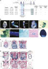
An essential CArG box in the miR-143/145 upstream enhancer. (A) Summary of transgenic constructs used to delineate the miR-143/145 enhancer is shown. Evolutionary conservation of the genomic region upstream of pre-miR-143 is shown at the top. Numbers of F0 transgenic embryos showing cardiac expression at E12.5 per total transgenic embryos obtained are indicated in the right column. A single mutation of the CArG in Reporter A abolished expression of the lacZ transgene in the embryonic heart and adult aorta. Embryonic heart expression was assayed in F0 and stable mouse lines, whereas adult aorta expression was assayed in two independent stable mouse lines. The 1.5-kb enhancer construct (Reporter B) contains only the two evolutionarily conserved regions and is sufficient to drive lacZ expression in the embryonic heart and adult aorta. Mutation of the CArG box in Reporter B abolished lacZ expression in the embryonic heart and adult aorta as determined in two stable mouse lines. (B) Transgenic mice were generated with a lacZ reporter linked to 5.5 kb of genomic DNA upstream of the miR-143/145 gene (Reporter A in A). The embryo in panel e was cleared to allow visualization of internal structures. Panel f is a high magnification of the embryo in panel e to show lacZ expression in SMCs lining the dorsal aorta (ao) and intersomitic arteries (isa). (Panel g) Adult aorta. (Panel h) Subdermal vasculature. (Panel i) Transverse section showing lacZ staining in an artery in skeletal muscle. (Panel j) Bladder. (ao) Aorta; (cc) cardiac crescent; (ht) heart. (C) Histological sections of Reporter B transgenic mice at various embryonic stages. (Bars: panel a, 100 μm; panels b,c, 200 μm. (a) Common atrial chamber; (bc) bulbus cordis; (la) left atrium; (lv) left ventricle; (ra) right atrium; (rv) right ventricle; (v) common ventricular chamber. (D) Histological sections of transgenic mice with wild-type genomic regions or the same regions with mutations in the CArG box at the indicated ages. Note that the CArG box mutation abolishes lacZ expression in the heart and vasculature. (a) Common atrial chamber; (ao) aorta; (bc) bulbus cordis; (la) left atrium; (lv) left ventricle; (ra) right atrium; (rv) right ventricle; (v) common ventricular chamber. Bars: panels a,b,g,h, 100 μm; panels c–f, 200 μm.
Figure 3.
Figure 3.
Targeting of the miR-143/145 cluster. (A) Schematic of miR-143/145 mutations. (B) Northern blots of RNA from bladders of duplicate mice of each genotype. Mutant mice used for these experiments had the neomyocin resistance gene removed by Cre-mediated recombination. Deletion of either miR-143 or miR-145 does not affect expression of the other member of the pair.
Figure 4.
Figure 4.
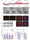
Smooth muscle abnormalities in miR-143/145 KO mice. (A) H&E staining of the dorsal aortae of mice of the indicated genotypes. Bar, 40 μm. (B) Electron microscopy of the dorsal aortae of mice of the indicated genotypes. Note the thickened ECM in miR-145 KO and dKO. Prominent actin-based stress fibers (arrows) are apparent in wild-type (WT) and miR-143 KO SMCs, but are absent from miR-145 KO and dKO SMCs. Rough endoplasmic reticulum (RER) is evident in miR-143 KO, miR-145 KO, and dKO SMCs, indicative of a synthetic state, but is not visible in wild-type SMCs. (ecm) Extracellular matrix; (enl) endothelial layer; (sf) stress fiber; (sm) smooth muscle. (C) Immunostaining of sections of aorta of wild-type and dKO mice showing similar staining for SMA. (D) Aortic SMCs were isolated from wild-type and dKO mice, cultured in growth medium, and stained for SMA. Wild-type SMCs display highly organized actin stress fibers, whereas SMCs from dKO mice display only diffuse actin staining. (E) Expression of the smooth muscle differentiation markers SMA and SM22 in aorta of wild-type and dKO mice as detected by Western blot analysis. GAPDH was detected as a loading control. (F) Expression of smooth muscle mRNAs as detected by real-time PCR. (G) Blood pressure measurements in wild-type and miR-145 KO mice. MAP and SBP were measured as described in the Materials and Methods. (*) P < 0.05.
Figure 5.
Figure 5.
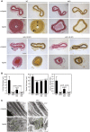
Reduced neointima formation in miR-143/145 mutant mice following carotid artery ligation. (A) Mice of the indicated genotypes were subjected to carotid artery ligation for 28 d, after which histological sections were obtained from the ligated artery and the unligated artery as a control 1900 μm proximal to the ligature and stained with H&E (left side) or for elastin (right side). The intimal (i) and medial (m) layers are designated with arrows in the ligated wild-type (WT) artery stained for elastin. Bar, 100 μm. (B) The intimal thickness in mice of each genotype 28 d after carotid artery ligation was determined by measuring the distance between the inner elastin layer and the lumen in at least five mice of each genotype. Medial thickness was measured as the distance between the inner and outer elastin layers. (C) Electron microscopy of carotid arteries of wild-type and dKO mice 14 d after ligation. SMCs from the wild-type artery adopt a migratory phenotype in response to ligation, giving rise to a neointima. In contrast, SMCs from the dKO artery retain a more organized arrangement and fail to form a neointima. The boundaries of representative SMCs are outlined in green. The dashed line in the bottom left panel indicates the internal boundary of the neointima.
Figure 6.
Figure 6.
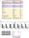
Modulation of actin dynamics by miR-143/145. (A) miR-143/145 targets involved in cytoskeletal remodeling and SRF activation. (B) The 3′ UTRs for the indicated mRNA targets of miR-143 and miR-145 were linked to luciferase and tested for repression by miR-143 and miR-145 expression plasmids in transfected COS cells. A miR-126 expression plasmid was used as a nonspecific (ns) control. The black bar (−) indicates the level of expression of the luciferase reporter without a cotransfected miRNA expression plasmid. miRNAs predicted to target each 3′ UTR are indicated at the top of each graph. (C) Aortae from mice of each genotype were isolated and protein lysates were analyzed for expression of KLF5 protein by Western blot analysis. GAPDH was included as a loading control. Numbers below each lane indicate relative expression of KLF5 normalized to GAPDH. (D) Carotid arteries were obtained from 10 wild-type and 10 dKO mice, pooled from each group, and expression of the indicated miR-143/145 targets was detected by real-time PCR.
Figure 7.
Figure 7.
Model for the role of miR-143 and miR-145 in the control of actin remodeling. MRTFs are sequestered in the cytoplasm by monomeric actin. Upon release from actin, MRTF translocates to the nucleus and interacts with SRF to activate the transcription of genes encoding actin and other cytoskeletal components, as well as miR-143 and miR-145. These miRNAs repress the expression of a collection of regulators of actin dynamics and MRTF/SRF activity, thereby creating a complex set of feedback loops to modulate cytoskeletal assembly and dynamics. (Adapted from Pipes et al. 2006.)

Similar articles

Cited by

References

    1. Barkalow KL, Italiano JE, Jr, Chou DE, Matsuoka Y, Bennett V, Hartwig JH. α-Adducin dissociates from F-actin and spectrin during platelet activation. J Cell Biol. 2003;161:557–570. - PMC - PubMed
    1. Barringhaus KG, Zamore PD. MicroRNAs: Regulating a change of heart. Circulation. 2009;119:2217–2224. - PMC - PubMed
    1. Bartel DP. MicroRNAs: Genomics, biogenesis, mechanism, and function. Cell. 2004;116:281–297. - PubMed
    1. Bonauer A, Carmona G, Iwasaki M, Mione M, Koyanagi M, Fischer A, Burchfield J, Fox H, Doebele C, Ohtani K, et al. MicroRNA-92a controls angiogenesis and functional recovery of ischemic tissues in mice. Science. 2009;324:1710–1713. - PubMed
    1. Chen J, Kitchen CM, Streb JW, Miano JM. Myocardin: A component of a molecular switch for smooth muscle differentiation. J Mol Cell Cardiol. 2002;34:1345–1356. - PubMed

Publication types

MeSH terms

LinkOut - more resources