. 2009 Nov 5;114(19):4310-9.
doi: 10.1182/blood-2009-03-211342. Epub 2009 Aug 28.

Bone marrow stem and progenitor cell contribution to neovasculogenesis is dependent on model system with SDF-1 as a permissive trigger

Affiliations

Bone marrow stem and progenitor cell contribution to neovasculogenesis is dependent on model system with SDF-1 as a permissive trigger

Gerard J Madlambayan et al. Blood. .

Abstract

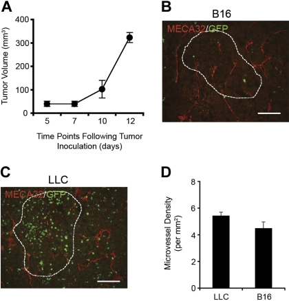
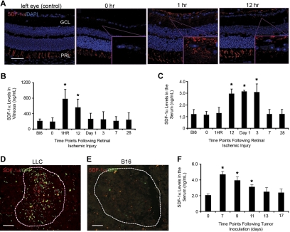
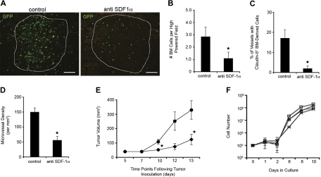
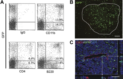
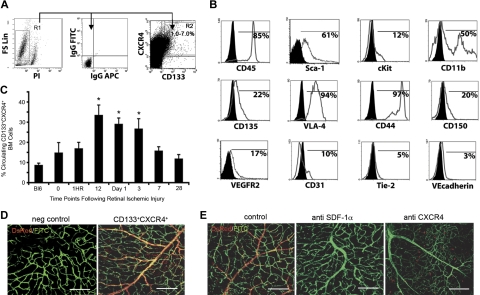
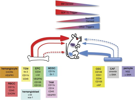
Adult bone marrow (BM) contributes to neovascularization in some but not all settings, and reasons for these discordant results have remained unexplored. We conducted novel comparative studies in which multiple neovascularization models were established in single mice to reduce variations in experimental methodology. In different combinations, BM contribution was detected in ischemic retinas and, to a lesser extent, Lewis lung carcinoma cells, whereas B16 melanomas showed little to no BM contribution. Using this spectrum of BM contribution, we demonstrate the necessity for site-specific expression of stromal-derived factor-1alpha (SDF-1alpha) and its mobilizing effects on BM. Blocking SDF-1alpha activity with neutralizing antibodies abrogated BM-derived neovascularization in lung cancer and retinopathy. Furthermore, secondary transplantation of single hematopoietic stem cells (HSCs) showed that HSCs are a long-term source of neovasculogenesis and that CD133(+)CXCR4(+) myeloid progenitor cells directly participate in new blood vessel formation in response to SDF-1alpha. The varied BM contribution seen in different model systems is suggestive of redundant mechanisms governing postnatal neovasculogenesis and provides an explanation for contradictory results observed in the field.


Figures

Figure 1
Figure 1
Differential BM contribution results from activation of redundant mechanisms of postnatal neovasculogenesis. Combinations of models of adult neovasculogenesis were established in single mice and result in different levels of BM contribution. (A-B) Lewis lung carcinoma cell (LLC)–based tumors showed GFP+ bone marrow (BM) contribution throughout the tumor mass (A; scale bar represents 100 μm) mainly from CD11b+ cells (B; n = 8; scale bar represents 50 μm). Tumors are outlined with dashed lines. (C-D) Within tumor-associated vasculature, CD31 (C) and claudin-5 (D) staining showed integration from GFP+ BM cells (scale bars represent 20 μm). (E) B16 tumors had low levels of GFP+ BM contribution in comparison with all other models and no contribution within tumor-associated vasculature (n = 8; scale bar represents 100 μm). (F) The most robust contribution was seen in the retinal injury model. This model uses vascular endothelial growth factor (VEGF) overexpression by a recombinant adeno-associated virus type 2 that overexpresses the murine 188 isoform of VEGF-A (rAAV2 VEGF-A 188) and laser-induced ischemic injury to promote robust BM-derived neovascularization. DsRed+ BM-derived blood vessels are shown (middle panel) along with a negative control (left panel; scale bars represent 100 μm). All animals were perfused with FITC-dextran to show functional vasculature (right panel; n = 5; scale bar represents 50 μm). Similar results were observed with GFP+ BM (data not shown). (G) Retinas were costained with α-SMA to confirm endothelial phenotype (n = 5; scale bar represents 50 μm). (H) LLC tumors established in mice that received a transplant of DsRed+ BM along with retinal injury also showed BM integration into tumor vasculature (n = 5; scale bar represents 20 μm). Confocal microscopy with 0.5-micron Z-step analysis was necessary to identify nucleated cells coexpressing donor GFP or DsRed and endothelial proteins (CD31).
Figure 2
Figure 2
B16 melanoma neovessels in the absence of BM contribution. (A) B16 tumors were capable of robust growth even without BM-derived neovascularization (n = 8). (B-C) MECA-32 staining demonstrated the presence of new blood vessels within B16 (B) and LLC tumors (C; scale bars represent 100 μm). (D) By quantifying MVD, it was found that B16 and LLC tumors contained microvessels at similar densities. LLC and B16 tumors are outlined with dashed lines.
Figure 3
Figure 3
Endogenously produced SDF-1α is a trigger for BM contribution to sites of postnatal neovasculogenesis. (A) Eyes were harvested and embedded in paraffin at different time points after retinal injury. Tissues were sectioned (5 μm) and staining was performed for SDF-1α and DAPI (4,6 diamidino-2-phenylindole). As expected, consistent expression of SDF-1α in the PRL was observed. Nontreated left eyes served as controls. SDF-1α expression is observed in the GCL at 1 and 12 hours after laser injury (n = 5; scale bar represents 100 μm). (B-C) SDF-1α ELISA of samples from the vitreous and blood serum after retinal ischemic injury. Vitreous fluid and blood serum were obtained from mice at different time points after retinal injury. ELISA analysis showed a direct correlation to the staining results with significant increases in SDF-1α levels observed at 1 hour and 12 hours in the vitreous (B) and blood serum (C), respectively (n = 5; *P < .05). (D-E) SDF-1α expression was also observed in LLC tumors (D) with nondetectable levels seen in B16 tumors (E). Tumors are outlined by dashed lines (n = 8; scale bars represent 100 μm). (F) ELISA analysis of SDF-1α in the serum of mice inoculated with both LLC and B16 tumors showed a significant increase in serum levels 7 days after tumor inoculation, with levels returning to background by day 13 (n = 5; *P < .05).
Figure 4
Figure 4
Blocking SDF-1α activity inhibits BM contribution to tumor neovascularization. After LLC inoculation, mice were treated with intratumoral anti–SDF-1α antibodies to block BM contribution (n = 5). (A-B) Tumors treated with anti–SDF-1α contained significantly lower numbers of BM-derived cells throughout the tumor mass seen visually (A; scale bars represent 100 μm) and by counting GFP+ cell numbers in standardized field sizes (B; *P < .05). (C-D) Treated mice also had significantly fewer cells integrated within blood vessel walls (C) and decreased MVD (D) compared with control tumors (*P < .05). (E) Animals treated with anti–SDF-1α antibodies (♦) also generated significantly smaller tumors in comparison with controls (●; *P < .05). (F) The growth kinetics of LLCs in culture with various doses of anti–SDF-1α antibodies (0 to 50 μg/mL) were similar over the range of concentrations tested. No cytotoxicity was observed at 0 (○), 12.5 (□), 25 (▵), and 50 μg/mL (x) of the anti–SDF-1α antibodies in growth media (n = 4).
Figure 5
Figure 5
Progeny of single HSCs participate in neovascularization. (A) Mice that received a transplant of GFP+ BM cells from primary donors initially engrafted with single HSCs demonstrate multilineage engraftment. Shown are representative flow cytometry plots demonstrating both myeloid (CD11b) and lymphoid (CD4 and B220) cell populations in engrafted secondary mice. Also shown is a representative isotype control. (B) In these mice, LLC tumors showed substantial HSC-derived GFP+ cell contribution throughout the tumor mass (n = 9; scale bar represents 100 μm). (C) Confocal micrograph of HSC-derived GFP+ cell in blood vessel wall coexpressing CD31. Orthogonal views created from confocal sections verify luminal expression of CD31 on HSC-derived cells.
Figure 6
Figure 6
BM-derived CD133+CXCR4+ cells enrich for neovascularization potential. The retinal injury model that shows the most robust BM contribution was used to identify BM-derived cells with direct neovasculogenic potential. (A) BM isolated from wild-type C57Bl/6 mice was analyzed for CD133 and CXCR4 expression. (B) Flow cytometric analysis of gated CD133+CXCR4+ cells shows an expression pattern that includes markers known to be found on BM-derived cells that participate in neovasculogenesis. CD133+CXCR4+ cells express endothelial cell surface markers such as VEGFR2, CD31, VE-cadherin, and tie 2. They also express CD45, CD117 (c-kit), Sca-1, VLA-4, CD11b, CD44, CD150, and CD135 (flt-3). (C) Kinetic analysis of CD133+CXCR4+ cell mobilization into the peripheral blood of mice after retinal injury showed an increase in cell number that correlated with SDF-1α levels in blood serum (n = 6; *P < .05). (D) Retinal flat mounts of injured eyes after induction of retinal ischemic injury that were adoptively transplanted with 106 BM-derived CD133+CXCR4+DsRed+ cells showed contribution to retinal neovascularization (n = 6). Untreated left eye (negative control) is also shown (scale bars represent 100 μm). Similar results were observed with GFP+ BM (data not shown; E) CD133+CXCR4+DsRed+ cell transplanted eyes that underwent induction of retinal ischemic injury with the added step of intravitreal injection with PBS containing an anti–SDF-1α– or anti–CXCR4-neutralizing antibody to a final concentration of 1 μg/mL are shown. Note the absence of newly formed vessels from CD133+CXCR4+DsRed+ BM cells under these conditions. As a control, treated eyes that underwent induction of retinal ischemic injury were treated with PBS containing an isotype-matched control antibody (n = 6; scale bars represent 100 μm). All animals were perfused with FITC-dextran to show functional vasculature.
Figure 7
Figure 7
Redundant mechanisms of postnatal neovascularization. Based on the studies outlined herein, we postulate that redundant mechanisms exist to achieve postnatal neovascularization. Within BM and other tissues including blood vessels, reside cells that are capable of participating in new blood vessel formation. Examples of these cell types and their phenotypes are provided. The extent of contribution is dependent on the model system. Some models including injured retinas (red circle) demonstrate robust BM contribution resulting in entire BM-derived blood vessels. Other models like LLC tumors (purple tumor) will incorporate BM cells into new vasculature, but the microenvironment of the tumor may also use other means of tumor neovessel formation. In this setting, redundant mechanisms act in concert to achieve new blood vessel growth. Finally, models such as B16 tumors (blue tumor) will have little to no BM contribution and rely mainly on non–BM-derived cells to generate new vessels. At sites of neovascularization, SDF-1α acts as a regulatory molecule necessary for BM recruitment and participation. Active sites that do not express SDF-1α are much less prone to BM involvement and undergo neovascularization via a non–BM-derived mechanism.

Similar articles

Cited by

References

    1. Asahara T, Murohara T, Sullivan A, et al. Isolation of putative progenitor endothelial cells for angiogenesis. Science. 1997;275(5302):964–967. - PubMed
    1. Duda DG, Cohen KS, Kozin SV, et al. Evidence for incorporation of bone marrow-derived endothelial cells into perfused blood vessels in tumors. Blood. 2006;107(7):2774–2776. - PMC - PubMed
    1. Grant MB, May WS, Caballero S, et al. Adult hematopoietic stem cells provide functional hemangioblast activity during retinal neovascularization. Nat Med. 2002;8(6):607–612. - PubMed
    1. Lyden D, Hattori K, Dias S, et al. Impaired recruitment of bone-marrow-derived endothelial and hematopoietic precursor cells blocks tumor angiogenesis and growth. Nat Med. 2001;7(11):1194–1201. - PubMed
    1. Nolan DJ, Ciarrocchi A, Mellick AS, et al. Bone marrow-derived endothelial progenitor cells are a major determinant of nascent tumor neovascularization. Genes Dev. 2007;21(12):1546–1558. - PMC - PubMed

Publication types

MeSH terms