

Blockade of T-lymphocyte KCa3.1 and Kv1.3 channels as novel immunosuppression strategy to prevent kidney allograft rejection
- PMID: 19715983
- PMCID: PMC3343637
- DOI: 10.1016/j.transproceed.2009.06.025
Blockade of T-lymphocyte KCa3.1 and Kv1.3 channels as novel immunosuppression strategy to prevent kidney allograft rejection
Abstract
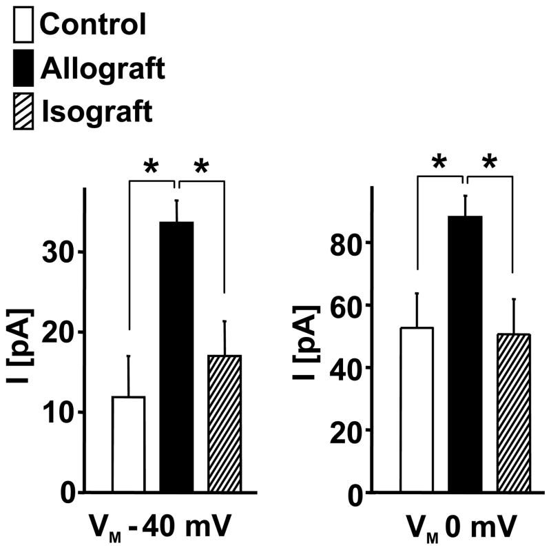
Currently, there is an unmet clinical need for novel immunosuppressive agents for long-term prevention of kidney transplant rejection as alternatives to the nephrotoxic calcineurin inhibitor cyclosporine (CsA). Recent studies have shown that K(+) channels have a crucial role in T-lymphocyte activity. We investigated whether combined blockade of the T-cell K(+) channels K(Ca)3.1 and K(v)1.3, both of which regulate calcium signaling during lymphocyte activation, is effective in prevention of rejection of kidney allografts from Fisher rats to Lewis rats. All recipients were initially treated with CsA (5 mg/kg d) for 7 days. In rats with intact allograft function, treatment was continued for 10 days with either CsA (5 mg/kg d), or a combination of TRAM-34 (K(Ca)3.1 inhibitor; 120 mg/kg d) plus Stichodactyla helianthus toxin (ShK, K(v)1.3 inhibitor; 80 microg/kg 3 times daily), or vehicle alone. Kidney sections were stained with periodic acid-Schiff or hematoxylin-eosin and histochemically for markers of macrophages (CD68), T-lymphocytes (CD43), or cytotoxic T-cells (CD8). Our results showed that treatment with TRAM-34 and ShK reduced total interstitial mononuclear cell infiltration (-42%) and the number of CD43+ T-cells (-32%), cytotoxic CD8+ T-cells (-32%), and CD68+ macrophages (-26%) in allografts when compared to vehicle treatment alone. Efficacy of TRAM-34/ShK treatment was comparable with that of CsA. In addition, no visible organ damage or other discernible adverse effects were observed with this treatment. Thus, selective blockade of T-lymphocyte K(Ca)3.1 and K(v)1.3 channels may represent a novel alternative therapy for prevention of kidney allograft rejection.
Figures


Similar articles
-
The Ca²⁺-activated K⁺ channel KCa3.1 as a potential new target for the prevention of allograft vasculopathy.PLoS One. 2013 Nov 29;8(11):e81006. doi: 10.1371/journal.pone.0081006. eCollection 2013. PLoS One. 2013. PMID: 24312257 Free PMC article.
-
Selective blockade of T lymphocyte K(+) channels ameliorates experimental autoimmune encephalomyelitis, a model for multiple sclerosis.Proc Natl Acad Sci U S A. 2001 Nov 20;98(24):13942-7. doi: 10.1073/pnas.241497298. Proc Natl Acad Sci U S A. 2001. PMID: 11717451 Free PMC article.
-
Blockage of K(Ca)3.1 and Kv1.3 channels of the B lymphocyte decreases the inflammatory monocyte chemotaxis.Int Immunopharmacol. 2016 Feb;31:266-71. doi: 10.1016/j.intimp.2015.12.032. Epub 2016 Feb 4. Int Immunopharmacol. 2016. PMID: 26795234
-
Potassium channel blockade by the sea anemone toxin ShK for the treatment of multiple sclerosis and other autoimmune diseases.Curr Med Chem. 2004 Dec;11(23):3041-52. doi: 10.2174/0929867043363947. Curr Med Chem. 2004. PMID: 15578998 Review.
-
K+ channel blockers: novel tools to inhibit T cell activation leading to specific immunosuppression.Curr Pharm Des. 2006;12(18):2199-220. doi: 10.2174/138161206777585120. Curr Pharm Des. 2006. PMID: 16787250 Review.
Cited by
-
Topical Delivery of Senicapoc Nanoliposomal Formulation for Ocular Surface Treatments.Int J Mol Sci. 2018 Sep 29;19(10):2977. doi: 10.3390/ijms19102977. Int J Mol Sci. 2018. PMID: 30274277 Free PMC article.
-
Physiological significance of delayed rectifier K(+) channels (Kv1.3) expressed in T lymphocytes and their pathological significance in chronic kidney disease.J Physiol Sci. 2015 Jan;65(1):25-35. doi: 10.1007/s12576-014-0331-x. Epub 2014 Aug 6. J Physiol Sci. 2015. PMID: 25096892 Free PMC article. Review.
-
T Cell Subset and Stimulation Strength-Dependent Modulation of T Cell Activation by Kv1.3 Blockers.PLoS One. 2017 Jan 20;12(1):e0170102. doi: 10.1371/journal.pone.0170102. eCollection 2017. PLoS One. 2017. PMID: 28107393 Free PMC article.
-
Immunomodulation by memantine in therapy of Alzheimer's disease is mediated through inhibition of Kv1.3 channels and T cell responsiveness.Oncotarget. 2016 Aug 16;7(33):53797-53807. doi: 10.18632/oncotarget.10777. Oncotarget. 2016. PMID: 27462773 Free PMC article.
-
The Ca²⁺-activated K⁺ channel KCa3.1 as a potential new target for the prevention of allograft vasculopathy.PLoS One. 2013 Nov 29;8(11):e81006. doi: 10.1371/journal.pone.0081006. eCollection 2013. PLoS One. 2013. PMID: 24312257 Free PMC article.
References
-
- Leichtman AB. Balancing efficacy and toxicity in kidney-transplant immunosuppression. N Engl J Med. 2007;357:2625. - PubMed
-
- Kahan BD. Cyclosporine. N Engl J Med. 1989;321:1725. - PubMed
-
- Mihatsch MJ, Kyo M, Morozumi K, et al. The side-effects of ciclosporine-A and tacrolimus. Clin Nephrol. 1998;49:356. - PubMed
Publication types
MeSH terms
Substances
Grants and funding
LinkOut - more resources
Full Text Sources
Other Literature Sources
Medical
Research Materials
