

Do plant cells secrete exosomes derived from multivesicular bodies?
- PMID: 19704795
- PMCID: PMC2633885
- DOI: 10.4161/psb.2.1.3596
Do plant cells secrete exosomes derived from multivesicular bodies?
Abstract
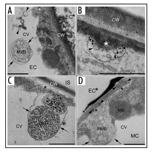
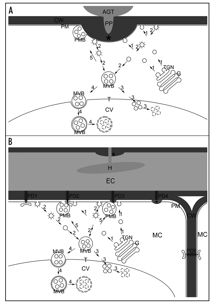
Multivesicular bodies (MVBs) are spherical endosomal organelles containing small vesicles formed by inward budding of the limiting membrane into the endosomal lumen. In mammalian red cells and cells of immune system, MVBs fuse with the plasma membrane in an exocytic manner, leading to release their contents including internal vesicles into the extracellular space. These released vesicles are termed exosomes. Transmission electron microscopy studies have shown that paramural vesicles situated between the plasma membrane and the cell wall occur in various cell wall-associated processes and are similar to exosomes both in location and in morphology. Our recent studies have revealed that MVBs and paramural vesicles proliferate when cell wall appositions are rapidly deposited beneath fungal penetration attempts or during plugging of plasmodesmata between hypersensitive cells and their intact neighboring cells. This indicates a potential secretion of exosome-like vesicles into the extracellular space by fusion of MVBs with the plasma membrane. This MVB-mediated secretion pathway was proposed on the basis of pioneer studies of MVBs and paramural vesicles in plants some forty years ago. Here, we recall the attention to the occurrence of MVB-mediated secretion of exosomes in plants.
Keywords: cell wall; endocytosis; endosome; exocytosis; exosome; multivesicular body; paramural body.
Figures
Comment on
-
Multivesicular compartments proliferate in susceptible and resistant MLA12-barley leaves in response to infection by the biotrophic powdery mildew fungus.New Phytol. 2006;172(3):563-76. doi: 10.1111/j.1469-8137.2006.01844.x. New Phytol. 2006. PMID: 17083686
-
Multivesicular bodies participate in a cell wall-associated defence response in barley leaves attacked by the pathogenic powdery mildew fungus.Cell Microbiol. 2006 Jun;8(6):1009-19. doi: 10.1111/j.1462-5822.2006.00683.x. Cell Microbiol. 2006. PMID: 16681841
Similar articles
-
Multivesicular bodies participate in a cell wall-associated defence response in barley leaves attacked by the pathogenic powdery mildew fungus.Cell Microbiol. 2006 Jun;8(6):1009-19. doi: 10.1111/j.1462-5822.2006.00683.x. Cell Microbiol. 2006. PMID: 16681841
-
Biogenesis and Function of Multivesicular Bodies in Plant Immunity.Front Plant Sci. 2018 Jul 9;9:979. doi: 10.3389/fpls.2018.00979. eCollection 2018. Front Plant Sci. 2018. PMID: 30038635 Free PMC article. Review.
-
Live-Imaging Detection of Multivesicular Body-Plasma Membrane Fusion and Exosome Release in Cultured Primary Neurons.Methods Mol Biol. 2023;2683:213-220. doi: 10.1007/978-1-0716-3287-1_17. Methods Mol Biol. 2023. PMID: 37300778
-
The Small GTPase Ral orchestrates MVB biogenesis and exosome secretion.Small GTPases. 2018 Nov 2;9(6):445-451. doi: 10.1080/21541248.2016.1251378. Epub 2016 Nov 22. Small GTPases. 2018. PMID: 27875100 Free PMC article.
-
Current knowledge on exosome biogenesis and release.Cell Mol Life Sci. 2018 Jan;75(2):193-208. doi: 10.1007/s00018-017-2595-9. Epub 2017 Jul 21. Cell Mol Life Sci. 2018. PMID: 28733901 Free PMC article. Review.
Cited by
-
Programmed cell death occurs asymmetrically during abscission in tomato.Plant Cell. 2011 Nov;23(11):4146-63. doi: 10.1105/tpc.111.092494. Epub 2011 Nov 29. Plant Cell. 2011. PMID: 22128123 Free PMC article.
-
Functional Nanomaterials for the Treatment of Osteoarthritis.Int J Nanomedicine. 2024 Jul 4;19:6731-6756. doi: 10.2147/IJN.S465243. eCollection 2024. Int J Nanomedicine. 2024. PMID: 38979531 Free PMC article. Review.
-
Tethering of Multi-Vesicular Bodies and the Tonoplast to the Plasma Membrane in Plants.Front Plant Sci. 2019 May 22;10:636. doi: 10.3389/fpls.2019.00636. eCollection 2019. Front Plant Sci. 2019. PMID: 31396242 Free PMC article.
-
Plant-derived extracellular vesicles: a novel nanomedicine approach with advantages and challenges.Cell Commun Signal. 2022 May 23;20(1):69. doi: 10.1186/s12964-022-00889-1. Cell Commun Signal. 2022. PMID: 35606749 Free PMC article. Review.
-
Structural Features of a Full-Length Ubiquitin Ligase Responsible for the Formation of Patches at the Plasma Membrane.Int J Mol Sci. 2021 Aug 31;22(17):9455. doi: 10.3390/ijms22179455. Int J Mol Sci. 2021. PMID: 34502365 Free PMC article.
References
-
- Gruenberg J, Stenmark H. The biogenesis of multivesicular endosomes. Nat Rev Mol Cell Biol. 2004;5:317–323. - PubMed
-
- Geldner N. The plant endosomal system-its structure and role in signal transduction and plant development. Planta. 2004;219:547–560. - PubMed
-
- Denzer K, Kleijmeer MJ, Heijnen HFG, Stoorvogel W, Geuze HJ. Exosome: From internal vesicle of the multivesicular body to intercellular signaling device. J Cell Sci. 2000;113:3365–3374. - PubMed
-
- An Q, Ehlers K, Kogel KH, van Bel AJE, Hückelhoven R. Multivesicular compartments proliferate in susceptible and resistant MLA12-barley leaves in response to infection by the biotrophic powdery mildew fungus. New Phytol. 2006;172:563–576. - PubMed
LinkOut - more resources
Full Text Sources
Other Literature Sources