. 2009 Nov;29(21):5696-709.
doi: 10.1128/MCB.00191-09. Epub 2009 Aug 24.

Protein phosphatase 2A-dependent dephosphorylation of replication protein A is required for the repair of DNA breaks induced by replication stress

Affiliations

Protein phosphatase 2A-dependent dephosphorylation of replication protein A is required for the repair of DNA breaks induced by replication stress

Junjie Feng et al. Mol Cell Biol. 2009 Nov.

Abstract

Eukaryotic genomic integrity is safeguarded by cell cycle checkpoints and DNA repair pathways, collectively known as the DNA damage response, wherein replication protein A (RPA) is a key regulator playing multiple critical roles. The genotoxic insult-induced phosphorylation of the 32-kDa subunit of human RPA (RPA32), most notably the ATM/ATR-dependent phosphorylation at T21 and S33, acts to suppress DNA replication and recruit other checkpoint/repair proteins to the DNA lesions. It is not clear, however, how the DNA damage-responsive function of phosphorylated RPA is attenuated and how the replication-associated activity of the unphosphorylated form of RPA is restored when cells start to resume the normal cell cycle. We report here that in cells recovering from hydroxyurea (HU)-induced genotoxic stress, RPA32 is dephosphorylated by the serine/threonine protein phosphatase 2A (PP2A). Interference with PP2A catalytic activity causes persistent RPA32 phosphorylation and increased HU sensitivity. The PP2A catalytic subunit binds to RPA following DNA damage and can dephosphorylate RPA32 in vitro. Cells expressing a RPA32 persistent phosphorylation mimetic exhibit normal checkpoint activation and reenter the cell cycle normally after recovery but display a pronounced defect in the repair of DNA breaks. These data indicate that PP2A-mediated RPA32 dephosphorylation is required for the efficient DNA damage repair.


Figures
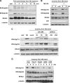
FIG. 1.
FIG. 1.
RPA32 undergoes dephosphorylation in cells recovering from HU block. (A) HeLa cells were treated with pulses of HU (0.2 mM) for 24 h, washed twice with PBS, and allowed to recover in drug-free medium. The levels of phosphorylated RPA32 were detected at the indicated recovery time points by immunoblotting. untr, untreated. (B and C) HeLa cells were released from HU block (0.2 mM, 24 h). The attenuation of RPA32 phosphorylation at T21/S33 was analyzed at 0 h or 9 h postrecovery in the absence (−) or presence (+) of MG132 (20 μM) (B) or different doses of OA (0 to 100 nM) or OA (50 nM) combined with caffeine (2 mM) (C). (D) A panel of five different cell lines (HeLa, HaCaT, HepG2, U2OS, and A549) were released from HU block (0.2 mM, 24 h) and then analyzed for RPA32 dephosphorylation at T21 at the indicated times after recovery.
FIG. 2.
FIG. 2.
PP2A mediates RPA32 dephosphorylation at T21 and S33. (A) HeLa cells were transfected with a mock siRNA oligonucleotide or with oligonucleotides against the catalytic subunits of PP1, PP2A, PP4, PP5, and PP6. Twenty-four hours posttransfection, cells were pulse treated with HU (0.2 mM, 24 h), and RPA32 dephosphorylation at T21/S33 was compared at the indicated time points. (B) HeLa cells transfected with control siRNA or siRNA against PP2A/C were pulse treated with HU (0.2 mM, 24 h). At 0 to 12 h postrelease, the cells were fixed and costained with DAPI and anti-RPA32pS33 and anti-γ-H2AX antibodies. The DNA damage-inducible foci were visualized by immunofluorescence microscopy, and the percentage of phospho-RPA32 and γ-H2AX focus-positive cells was determined, normalized, and plotted. The error bars represent the standard deviations from three independent experiments. (C) Immunoprecipitation (IP) assays were performed in unperturbed cells or cells recovering from exposure to pulses of HU (0.2 mM, 24 h) at the indicated times after release using an anti-RPA32 antibody. The association of RPA32 with PP2A was detected by immunoblotting (IB) using an antibody against PP2A/C (α-PP2A/C). Binding of RPA70 was also examined as a loading control. The positions of the immunoglobulin light and heavy chains detected in the reaction are indicated by asterisks. untr, untreated; IgG, immunoglobulin G. (D) HeLa cells were mock treated (untreated [untr]) or exposed to HU (0.2 mM) for 24 h. DNA damage-inducible foci of RPA32 and PP2A were examined by immunofluorescence. (E) RPA32 was immunoprecipitated from HeLa cells exposed to HU (2 mM) for 24 h and used as substrate for the in vitro phosphatase assay in the absence (−) or presence (+) of purified PP2A catalytic subunit and different doses of OA (0 to 25 nM). Western blot analysis was performed 30 min later to determine the level of phosphorylated RPA32.
FIG. 3.
FIG. 3.
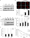
RPA32 dephosphorylation at T21/S33 is dispensable for the checkpoint activation but is required for recovery from HU stress. (A) The various cell lines with substitutions in RPA32 (S) were created by stably expressing in HeLa cells the empty vector (V) or Flag-tagged RPA32 variants (WT, VA, and DD) followed by retrovirally silencing endogenous RPA32 in these overexpression cells (O). RPA32 levels in these cell lines were examined by Western blot analysis. (B) WT and DD cells were treated with HU (5 mM). At 0 to 3 h postexposure, cells were fixed and stained with anti-RPA32 antibody. The RPA32 foci were visualized by immunofluorescence, and the percentage of focus-positive cells was determined, normalized, and plotted. Error bars represent the standard deviations from three independent experiments. (C) The RPA32 substitution cells were mock treated (−) or exposed (+) to HU (5 mM), and phosphorylation of Chk1 at S345 and Rad17 at S645 was examined by Western blotting 3 h later. (D) Various cells were irradiated with 25 J/m2 UV, and 3 h later, the DNA synthesis rate was determined and normalized to that in untreated cells (Untr). (E) The RPA32 substitution cells were left untreated or irradiated with 25 J/m2 UV. Later (1.5 h later), the cells were costained with propidium iodide and anti-phospho-H3 antibodies, and the mitotic fractions were determined by fluorescence-activated cell sorting analysis and normalized to that of unperturbed cells. (F) The RPA32 substitution cell lines were pulse exposed to 0 to 1.2 mM HU for 24 h and then allowed to recover in drug-free medium for 10 to 14 days. The cell viability of various lineages was analyzed by the clonogenic survival assay. All the data points in panels D, E, and F represent the means ± standard deviations (error bars) from three independent experiments, each performed in triplicate.
FIG. 4.
FIG. 4.
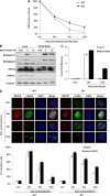
RPA32 dephosphorylation is not required for the resumption of DNA replication or subsequent mitosis following release from HU block. (A) The RPA32 substitution cells were released from HU exposure (pulses) (0.2 mM, 24 h). At 0 to 3 h postrelease, the DNA synthesis rates were determined and normalized to the rate at 0-h time point. All samples were tested in triplicate, and consistent results were obtained among three independent experiments. (B) Various cell lineages were allowed to recover from pulse exposure to HU (0.2 mM, 24 h). Cells at the indicated time points were fixed and costained with propidium iodide (PI) and anti-phospho-H3 antibody, and the mitotic fractions were further determined by fluorescence-activated cell sorting (FACS) analysis. (C and D) HeLa cells were treated with sublethal (0.2 mM) or lethal (2 mM) doses of HU for 24 h and then allowed to recover. At 0 to 24 h after release, cells were harvested and analyzed by immunoblotting using appropriate antibodies (C). The remaining cells underwent cell cycle profiling analysis by PI staining and FACS (D). (E) HeLa cells were pulse exposed to lethal doses of HU (2 mM, 24 h). At 6 h postrecovery, cells were pulse-labeled with BrdU for 10 min followed by immunofluorescence analysis using anti-BrdU and anti-RPA32pS33 antibodies. The cell positive for both types of staining were boxed and magnified for clearer visualization. (F) HeLa cells were allowed to recover from HU treatment (0.2 or 2 mM, 24 h) (pulses), and mitotic index analysis was performed by FACS at the indicated time points after release.
FIG. 5.
FIG. 5.
RPA32 dephosphorylation is necessary for the efficient repair of DNA breaks induced by HU. (A) WT and DD cells were allowed to recover from HU exposure (0.2 mM, 24 h) (pulses). At 0 to 12 h postrecovery, cells were harvested and analyzed by alkaline comet assay. Repair of DNA breaks was evaluated by the level of the residual DNA breaks, which is calculated by the comet tail moments at various recovery time points relative to that at the 0-h time point. (B and C) HeLa cells were transfected with control siRNA or siRNA against the PP2A catalytic subunit. At 24 h posttransfection, the cells were pulse exposed to HU (0.2 mM, 24 h) and then allowed to recover. RPA32 dephosphorylation was analyzed by immunoblotting (B), and repair of DNA breaks was evaluated by alkaline comet assay (C). untr, untreated; RNAi, RNA interference. (D) WT and DD cells were allowed to recover from HU treatment (0.2 mM, 24 h) (pulses). At 0 to 12 h postrelease, cells were fixed and stained with DAPI and anti-RPA70 and anti-γ-H2AX antibodies. Immunofluorescence was carried out to visualize the DNA damage foci formed by RPA70 and γ-H2AX, and the percentages of the focus-positive cells were calculated, normalized, and plotted. All the data points in panels A, C, and D represent the means ± standard deviations (error bars) from three independent experiments.

Similar articles

Cited by

References

    1. Abraham, R. T. 2001. Cell cycle checkpoint signaling through the ATM and ATR kinases. Genes Dev. 15:2177-2196. - PubMed
    1. Ali, A., J. Zhang, S. Bao, I. Liu, D. Otterness, N. M. Dean, R. T. Abraham, and X. F. Wang. 2004. Requirement of protein phosphatase 5 in DNA-damage-induced ATM activation. Genes Dev. 18:249-254. - PMC - PubMed
    1. Arnold, H. K., and R. C. Sears. 2006. Protein phosphatase 2A regulatory subunit B56β associates with c-Myc and negatively regulates c-Myc accumulation. Mol. Cell. Biol. 26:2832-2844. - PMC - PubMed
    1. Bartek, J., and J. Lukas. 2007. DNA damage checkpoints: from initiation to recovery or adaptation. Curr. Opin. Cell Biol. 19:238-245. - PubMed
    1. Binz, S. K., Y. Lao, D. F. Lowry, and M. S. Wold. 2003. The phosphorylation domain of the 32-kDa subunit of replication protein A (RPA) modulates RPA-DNA interactions. Evidence for an intersubunit interaction. J. Biol. Chem. 278:35584-35591. - PubMed

Publication types

MeSH terms

LinkOut - more resources