. 2009 Sep;16(9):945-52.
doi: 10.1038/nsmb.1648. Epub 2009 Aug 16.

A molecular basis for phosphorylation-dependent SUMO conjugation by the E2 UBC9

Affiliations

A molecular basis for phosphorylation-dependent SUMO conjugation by the E2 UBC9

Firaz Mohideen et al. Nat Struct Mol Biol. 2009 Sep.

Abstract

Phosphorylation and small ubiquitin-like modifier (SUMO) conjugation contribute to the spatial and temporal regulation of substrates containing phosphorylation-dependent SUMO consensus motifs (PDSMs). Myocyte-enhancement factor 2 (MEF2) is a transcription factor and PDSM substrate whose modification by SUMO drives postsynaptic dendritic differentiation. NMR analysis revealed that the human SUMO E2 interacted with model substrates for phosphorylated and nonphosphorylated MEF2 in similar extended conformations. Mutational and biochemical analysis identified a basic E2 surface that enhanced SUMO conjugation to phosphorylated PDSM substrates MEF2 and heat-shock transcription factor 1 (HSF1), but not to nonphosphorylated MEF2 or HSF1, nor the non-PDSM substrate p53. Mutant ubiquitin-conjugating enzyme UBC9 isoforms defective in promoting SUMO conjugation to phosphorylated MEF2 in vitro and in vivo also impair postsynaptic differentiation in organotypic cerebellar slices. These data support an E2-dependent mechanism that underlies phosphorylation-dependent SUMO conjugation in pathways that range from the heat-shock response to nuclear hormone signaling to brain development.


Figures

Figure 1
Figure 1
Phosphorylation-dependent SUMO conjugation is mediated by the SUMO E2. (a) Extended SUMO consensus motif highlighting the substrate lysine and the phosphorylated serine residues. (b) Sequence alignment for 20 amino acids encompassing the Phosphorylation-dependent SUMO conjugation motif (PDSM) from human MEF2 family members, HSF1, HSF4, GATA-1, PPARγ, the Negatively charged amino acid dependent SUMO conjugation motif (NDSM) from human Elk1 and 15 amino acids encompassing the p53 SUMO consensus motif. The amino acid number for each C-terminal residue is indicated in parentheses. (c) Upper panel; insets showing examples of SDS-PAGE analysis and fluorescent detection for SUMO conjugation to MEF2P and MEF2 at substrate concentrations of 80 μM. Additional examples for a subset of raw data used in constructing plots in the kinetic analysis provided in Supplementary Figures 1 and 2. Lower panel; plot shown on the left for initial rates of reaction versus substrate concentration. The bar chart to the right of the plot indicates individual specificity constants (k2/Kd) for wild-type Ubc9 mediated SUMO conjugation to MEF2P (red) and MEF2 (black). The bar chart (right) indicates the fold preference for MEF2P (gray) expressed as MEF2P(k2/Kd)/MEF2(k2/Kd). (d) HA-SUMO1 conjugation to wild-type GAL4-MEF2A and GAL4-MEF2A-S408A in 293T cells. Left panel; GAL4 immunoprecipitates of 293T cells transfected with HA-SUMO1 and wild-type, K403R or S408A mutant GAL4-MEF2A were immunoblotted using the indicated antibodies. Immunoblot of total lysate with anti-tubulin antibody (as loading control). Right panel; bar chart indicating 4.08 ± 1.48 enhancement for HA-SUMO1 modification to wild-type GAL4-MEF2A compared to GAL4-MEF2A-S408A calculated as described in Methods. Biochemical assays were conducted in triplicate. Error bars are ±1 standard deviation.
Figure 2
Figure 2
Model for PDSM recognition by the E2 Ubc9. (a) Surface representation and electrostatic potential of Ubc9 proximal to the RanGAP SUMO consensus motif (ψ-K-x-E SUMO consensus motif obtained from a previously characterized complex between RanGAP and Ubc9; PDB ID 2GRN). The positions for three C-terminal amino acids including the phosphorylated serine residue were obtained from a previously characterized complex between Pin1 and a phosphorylated peptide (PDB ID 1F8A) and combined with the RanGAP SUMO consensus sequence to model the PDSM. The relevant amino acids discussed in the text are labeled using the three-letter code on or adjacent to their position on the Ubc9 surface. Blue represents basic surfaces while red represents acidic surfaces. (b) Model for the Ubc9:PDSM complex. The structure of Ubc9 is shown in ribbon representation and the PDSM residues in stick representation. The SUMO consensus motif and the three C-terminal residues were obtained as in a. All structures graphically depicted using PyMol.
Figure 3
Figure 3
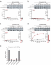
Kinetic and mutational analyses for Ubc9 amino acid residues involved in PDSM discrimination. (a) Upper panel; insets showing examples of SDS-PAGE analysis and fluorescent detection for Ubc9-K65A mediated SUMO conjugation to MEF2P and MEF2 at substrate concentrations of 80 μM. Lower panel; the plot on the left indicates initial rates versus substrate concentration and the bar chart shown on the right indicates the specificity constants (k2/Kd) for Ubc9-K65A mediated SUMO conjugation to MEF2P (red) and MEF2 (black). (b), (c) and (d) Similar data as in a, but for Ubc9-K74A, Ubc9-K76A and Ubc9-K59A respectively. (e) The bar chart indicates the fold preference for MEF2P (gray) expressed as MEF2P(k2/Kd)/MEF2(k2/Kd) for wild-type Ubc9, Ubc9-K65A, Ubc9-K74A, Ubc9-K76A and Ubc9-K59A. The dashed red line indicates an equal preference for Ubc9-mediated SUMO conjugation to MEF2P and MEF2. Biochemical assays were conducted in triplicate. Error bars are ±1 standard deviation.
Figure 4
Figure 4
Ubc9 Lys65 is important for PDSM discrimination of HSF1 but not for the non-PDSM substrate p53. (a) Upper panel; insets showing examples of SDS-PAGE analysis and fluorescent detection for wild-type Ubc9 mediated SUMO conjugation to HSF1P and HSF1 at substrate concentrations of 80 μM. Lower panel; the plot on the left indicates initial rates versus substrate concentration and the bar chart shown on the right indicates the specificity constants (k2/Kd) for wild-type Ubc9 mediated SUMO conjugation to HSF1P (red) and HSF1 (black). (b) Similar data as in a, but for Ubc9-K65A. (c) Upper panel; insets showing examples of SDS-PAGE analysis and fluorescent detection for wild-type Ubc9 mediated SUMO conjugation to p53 at a substrate concentrations of 16 μM. Lower panel; the plot on the left indicates initial rates versus substrate concentration and the bar chart shown on the right indicates the specificity constant (k2/Kd) for wild-type Ubc9 mediated SUMO conjugation to p53. (d) Similar data as in c, but for Ubc9-K65A. Biochemical assays were conducted in triplicate. Error bars are ±1 standard deviation.
Figure 5
Figure 5
Amino acid side chains that constitute the basic surface on Ubc9 are important for PDSM discrimination of MEF2 in vivo. (a) SDS-PAGE analysis of whole-cell lysate obtained from 293T cells transfected with HA-SUMO1 (left panel) or HA-SUMO2 (right panel) and wild-type Myc-UBC9 or the indicated mutant isoforms immunoblotted using anti-HA, anti-Myc, or anti-tubulin antibodies (loading control). (b) Upper panel; SDS-PAGE analysis of GAL4 immunoprecipitates from 293T cells transfected with HA-SUMO1, indicated Myc-UBC9 alleles and GAL4-MEF2A immunoblotted with anti-HA and anti-GAL4 antibodies as well as anti-myc and anti-tubulin antibodies (loading controls). Lower panel; bar chart indicating ratios between the preference of SUMO modification for wild-type GAL4-MEF2A and GAL4-MEF2-S408A for wild-type and mutant Ubc9 isoforms obtained from three independent transfection (Methods) except for Myc-Ubc9-K59A which was calculated from two independent experiments due to technical difficulties. Ratios for SUMO conjugation (GAL4-MEF2A/GAL4-MEF2A-S408A) were 9.40 ± 3.67, 1.24 ± 0.35, 0.78 ± 0.43, 2.03 ± 1.39 and 8.30 ± 1.89 for Myc-Ubc9, Myc-Ubc9-K65A, Myc-Ubc9-K74A, Myc-Ubc9-K76A and Myc-Ubc9-K59A, respectively. Biochemical assays were conducted in triplicate. Error bars are ±1 standard deviation. (c) SUMO2 conjugation to MEF2A in 293T cells. GAL4 immunoprecipitates of 293T cells transfected with HA-SUMO2 and the indicated mutant isoforms of Myc-UBC9 and GAL4-MEF2A were immunoblotted with HA and GAL4 antibodies. Total lysates were immunoblotted with a myc antibody for Myc-Ubc9 and a tubulin antibody which served as a loading control for the SDS-PAGE analysis.
Figure 6
Figure 6
Ubc9 mutants deficient in PDSM discrimination are also deficient for dendritic claw differentiation in the cerebellar cortex. (a) Representative confocal images of GFP-positive granule neurons expressing vector (left), Flag-Ubc9-WT (middle), or Flag-Ubc9-K65A (right). Arrows and arrowheads indicate dendritic claws and axons, respectively. Insets show higher-magnification views of ends of dendrites. Main scale bar is 20 microns; inset scale bar is 4 microns. Asterisk denotes an axon of a neuron that is not pictured. (b) Quantification of the effect of Ubc9 and Ubc9 mutants on dendritic claw differentiation in rat cerebellar slices. The number of dendritic claws was significantly reduced in granule neurons expressing Flag-Ubc9-K65A, Flag-Ubc9-K74A, and Flag-Ubc9-K76A as compared to those cells expressing Flag-Ubc9 WT-expressing neurons (p<0.001; ANOVA, followed by Bonferroni-Dunn post-hoc test). Vector, Flag-Ubc9-WT, and Flag-Ubc9-K59A expressing granule neurons were not markedly different from each other. (c) SDS-PAGE analysis and immunoblots using lysates obtained from 293T cells transfected with expression plasmids encoding Flag-UBC9 WT or indicated UBC9 mutant alleles. Error bars are ±1 standard deviation.

Similar articles

Cited by

References

    1. Johnson ES. Protein modification by SUMO. Annu. Rev. Biochem. 2004;73:355–382. - PubMed
    1. Geiss-Freidlander R, Melchior F. Concepts in sumoylation: a decade on. Nat. Rev. Mol. Cell Biol. 2007;8:947–56. - PubMed
    1. Dasso M. Emerging roles of the SUMO pathway in mitosis. Cell Div. 2008;3:5. - PMC - PubMed
    1. Hershko A, Ciechanover A. The ubiquitin system. Annu. Rev. Biochem. 1998;67:425–479. - PubMed
    1. Dye BT, Schulman BA. Structural mechanism underlying posttranslational modification by ubiquitin-like proteins. Annu. Rev. Biophys. Biomol. Struct. 2007;36:131–150. - PubMed

Publication types

MeSH terms

Substances