Uncoupling of astrogliosis from epileptogenesis in adenosine kinase (ADK) transgenic mice
- PMID: 19674507
- PMCID: PMC3045053
- DOI: 10.1017/S1740925X09990135
Uncoupling of astrogliosis from epileptogenesis in adenosine kinase (ADK) transgenic mice
Abstract
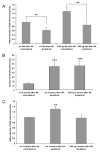
The astrocytic enzyme adenosine kinase (ADK) is a key negative regulator of the brain's endogenous anticonvulsant adenosine. Astrogliosis with concomitant upregulation of ADK is part of the epileptogenic cascade and contributes to seizure generation. To molecularly dissect the respective roles of astrogliosis and ADK-expression for seizure generation, we used a transgenic approach to uncouple ADK-expression from astrogliosis: in Adk-tg mice the endogenous Adk-gene was deleted and replaced by a ubiquitously expressed Adk-transgene with novel ectopic expression in pyramidal neurons, resulting in spontaneous seizures. Here, we followed a unique approach to selectively injure the CA3 of these Adk-tg mice. Using this strategy, we had the opportunity to study astrogliosis and epileptogenesis in the absence of the endogenous astrocytic Adk-gene. After triggering epileptogenesis we demonstrate astrogliosis without upregulation of ADK, but lack of seizures, whereas matching wild-type animals developed astrogliosis with upregulation of ADK and spontaneous recurrent seizures. By uncoupling ADK-expression from astrogliosis, we demonstrate that global expression levels of ADK rather than astrogliosis per se contribute to seizure generation.
Figures


Similar articles
-
Astrogliosis in epilepsy leads to overexpression of adenosine kinase, resulting in seizure aggravation.Brain. 2005 Oct;128(Pt 10):2383-95. doi: 10.1093/brain/awh555. Epub 2005 Jun 1. Brain. 2005. PMID: 15930047
-
Adenosine kinase is a target for the prediction and prevention of epileptogenesis in mice.J Clin Invest. 2008 Feb;118(2):571-82. doi: 10.1172/JCI33737. J Clin Invest. 2008. PMID: 18172552 Free PMC article.
-
Local disruption of glial adenosine homeostasis in mice associates with focal electrographic seizures: a first step in epileptogenesis?Glia. 2012 Jan;60(1):83-95. doi: 10.1002/glia.21250. Epub 2011 Sep 30. Glia. 2012. PMID: 21964979 Free PMC article.
-
The adenosine kinase hypothesis of epileptogenesis.Prog Neurobiol. 2008 Mar;84(3):249-62. doi: 10.1016/j.pneurobio.2007.12.002. Epub 2007 Dec 23. Prog Neurobiol. 2008. PMID: 18249058 Free PMC article. Review.
-
Adenosine dysfunction in epilepsy.Glia. 2012 Aug;60(8):1234-43. doi: 10.1002/glia.22285. Epub 2011 Dec 22. Glia. 2012. PMID: 22700220 Free PMC article. Review.
Cited by
-
Adenosine kinase: exploitation for therapeutic gain.Pharmacol Rev. 2013 Apr 16;65(3):906-43. doi: 10.1124/pr.112.006361. Print 2013 Jul. Pharmacol Rev. 2013. PMID: 23592612 Free PMC article. Review.
-
Inhibitory RNA in epilepsy: research tools and therapeutic perspectives.Epilepsia. 2010 Sep;51(9):1659-68. doi: 10.1111/j.1528-1167.2010.02672.x. Epub 2010 Jul 15. Epilepsia. 2010. PMID: 20633035 Free PMC article. Review.
-
Adenosine receptors and epilepsy: current evidence and future potential.Int Rev Neurobiol. 2014;119:233-55. doi: 10.1016/B978-0-12-801022-8.00011-8. Int Rev Neurobiol. 2014. PMID: 25175969 Free PMC article. Review.
-
Genetic variations of adenosine kinase as predictable biomarkers of efficacy of vagus nerve stimulation in patients with pharmacoresistant epilepsy.J Neurosurg. 2021 Sep 3;136(3):726-735. doi: 10.3171/2021.3.JNS21141. Print 2022 Mar 1. J Neurosurg. 2021. PMID: 34479194 Free PMC article.
-
Overexpression of ADK in human astrocytic tumors and peritumoral tissue is related to tumor-associated epilepsy.Epilepsia. 2012 Jan;53(1):58-66. doi: 10.1111/j.1528-1167.2011.03306.x. Epub 2011 Nov 16. Epilepsia. 2012. PMID: 22092111 Free PMC article.
References
-
- Bjorklund O, Shang MM, Tonazzini I, Dare E, Fredholm BB. Adenosine A(1) and A(3) receptors protect astrocytes from hypoxic damage. European Journal of Pharmacology. 2008;596:6–13. - PubMed
-
- Boison D. Adenosine kinase, epilepsy and stroke: mechanisms and therapies. Trends Pharmacol Sci. 2006;27:652–658. - PubMed
-
- Boison D. Adenosine-based cell therapy approaches for pharmacoresistant epilepsies. Neurodegener Dis. 2007;4:28–33. - PubMed
-
- Boison D. Astrogliosis and adenosine kinase: a glial basis of epilepsy. Future Neurology. 2008b;3:221–224.
Publication types
MeSH terms
Substances
Grants and funding
LinkOut - more resources
Full Text Sources
Medical
Miscellaneous

