. 2009 Aug 7;138(3):592-603.
doi: 10.1016/j.cell.2009.07.011.

Downregulation of miRNA-200c links breast cancer stem cells with normal stem cells

Affiliations

Downregulation of miRNA-200c links breast cancer stem cells with normal stem cells

Yohei Shimono et al. Cell. .

Abstract

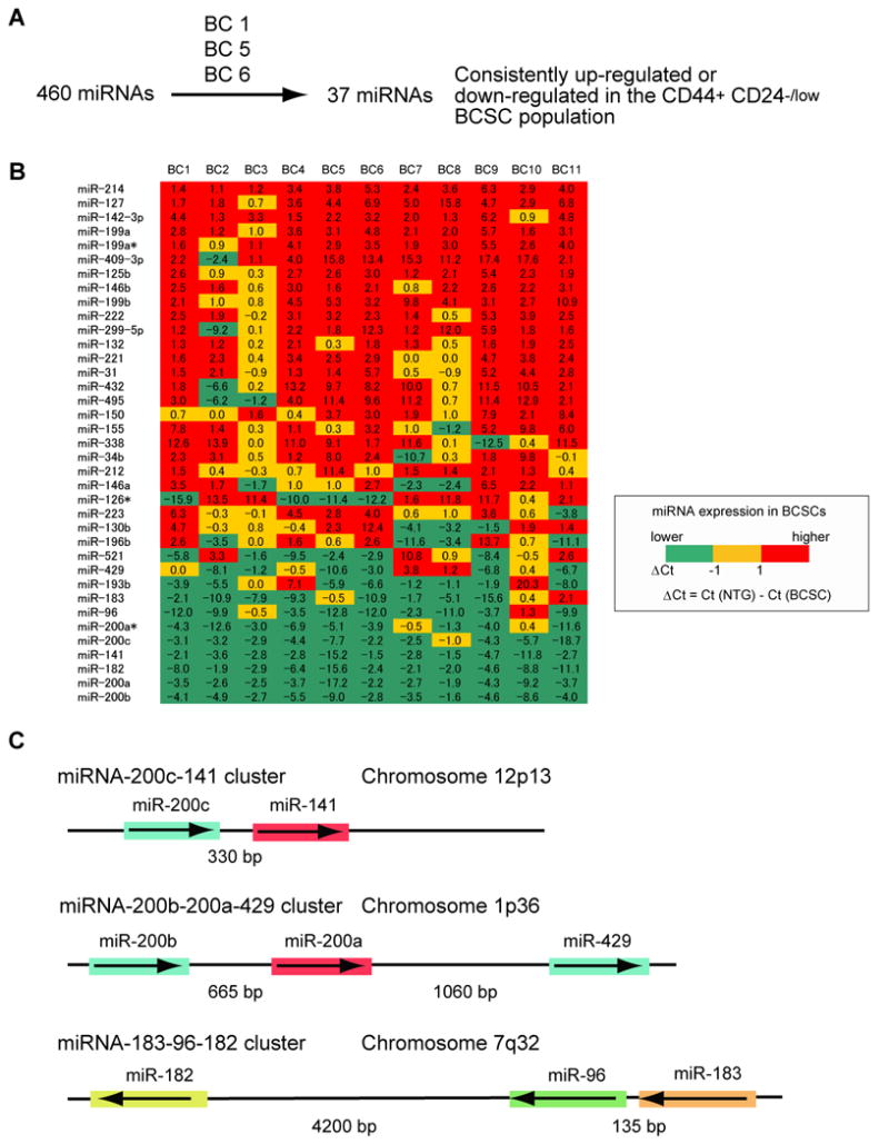
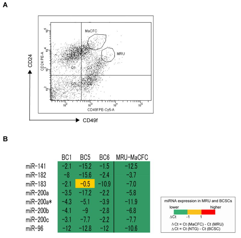
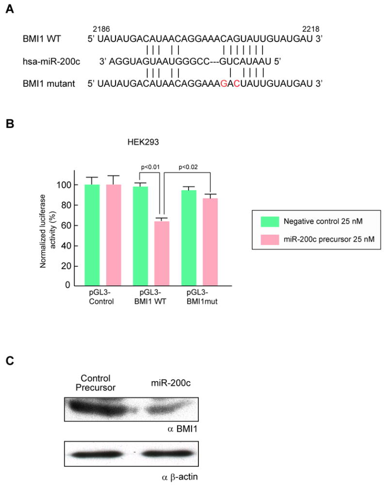
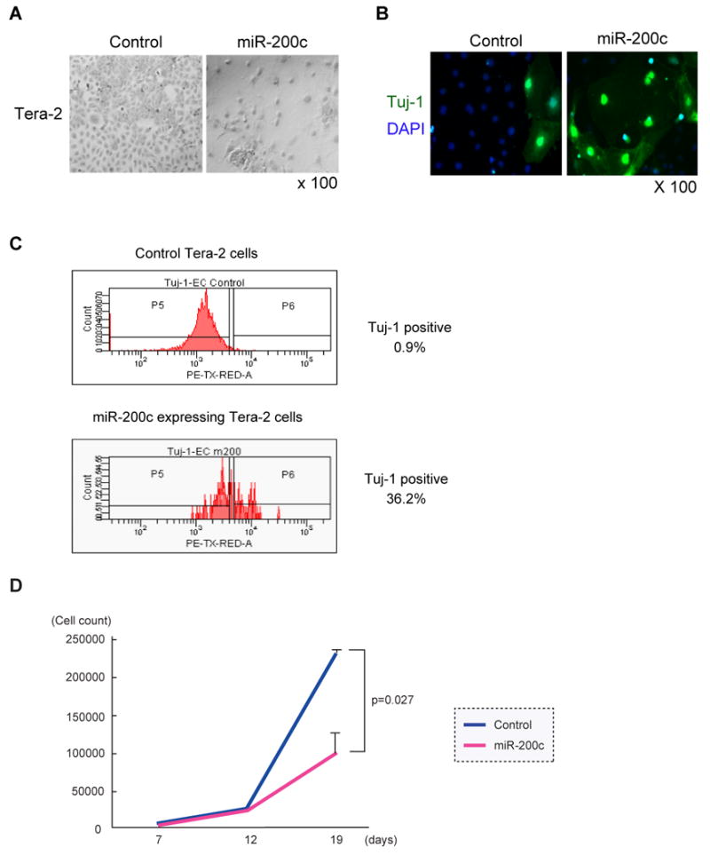
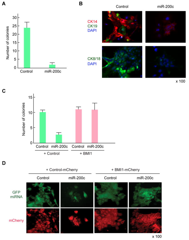
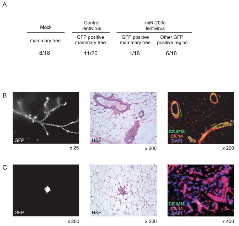
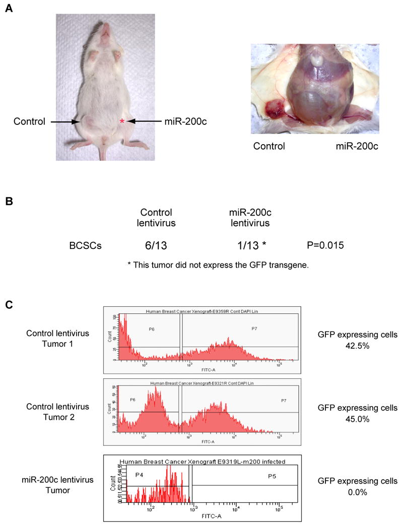
Human breast tumors contain a breast cancer stem cell (BCSC) population with properties reminiscent of normal stem cells. We found 37 microRNAs that were differentially expressed between human BCSCs and nontumorigenic cancer cells. Three clusters, miR-200c-141, miR-200b-200a-429, and miR-183-96-182 were downregulated in human BCSCs, normal human and murine mammary stem/progenitor cells, and embryonal carcinoma cells. Expression of BMI1, a known regulator of stem cell self-renewal, was modulated by miR-200c. miR-200c inhibited the clonal expansion of breast cancer cells and suppressed the growth of embryonal carcinoma cells in vitro. Most importantly, miR-200c strongly suppressed the ability of normal mammary stem cells to form mammary ducts and tumor formation driven by human BCSCs in vivo. The coordinated downregulation of three microRNA clusters and the similar functional regulation of clonal expansion by miR-200c provide a molecular link that connects BCSCs with normal stem cells.


Figures

Figure 1
Figure 1. Profile of Human Breast Cancer Stem Cell miRNA Expression
(A) Screening of 460 miRNA expression in human breast cancer stem cells. The details of the screen used to identify the 37 miRNAs differentially expressed by the CD44+CD24-/lowlineage- human breast cancer stem cells (BCSCs) and the remaining lineage- non-tumorigenic cancer cells (NTG cells) are shown schematically. (B) Expression profile of 37 miRNAs in human breast cancer stem cells. Flow cytometry was used to isolate BCSCs and NTG cells from 11 human breast cancer samples (BC1 to BC11). The amount of miRNA expression (Ct value) in 100 sorted cancer cells was analyzed by multiplex quantitative real-time PCR. Numbers represent the difference of Ct values (ΔCt) obtained from BCSCs and NTG cells. (C) A schematic representation of the three miRNA clusters down-regulated in human breast cancer stem cells. The miRNAs sharing the same seed sequence (from 2 to 7 base pairs) are marked by the same color.
Figure 2
Figure 2. Profile of Down-regulated miRNAs Shared Between Normal Mammary Stem Cells and Breast Cancer Stem Cells
(A) Distribution of CD45-CD31-CD140a-Ter119- mouse mammary cells according to their expression of CD24 and CD49f MRU is a population enriched for mammary stem cells. MaCFCs are enriched for progenitors that do not regenerate a mammary duct in vivo. (B) Expression of miRNAs in MRUs as compared to MaCFCs. The expression of the miRNAs down-regulated in human breast cancer stem cells was analyzed in MRUs and MaCFCs isolated by flow cytometry from normal mouse mammary fat pads. The level of miRNA expression in 100 MRUs or MaCFCs was measured by quantitative real-time PCR. The analysis was repeated twice by using the two sets of samples derived from independently isolated populations of MRUs and MaCFCs. Numbers represent the difference of Ct values obtained from MRUs and MaCFCs. BC1, BC5 and BC6 represent miRNA expression of human breast cancer stem cells described in Figure 1.
Figure 3
Figure 3. MiR-200c Targets BMI1
(A) Schematic representation of the miR-200bc/429 target sequence within the 3′ UTR of BMI1. Two nucleotides (complementary to nucleotide 6 and 8 of miR-200bc/429) were mutated in the 3′UTR of BMI1. The numbers indicate the position of the nucleotides in the reference wild-type sequences (NM_005180). (B) Activity of the luciferase gene linked to the 3′UTR of BMI1. The pGL3 firefly luciferase reporter plasmids with the wild-type or mutated 3′ UTR sequences of BMI1 were transiently transfected into HEK293 cells along with 25 nM miR-200c precursor or negative control, and a Renilla luciferase reporter for normalization. Luciferase activities were measured after 48 hours. The mean of the results from the cells transfected by pGL3 control vector was set as 100 %. The data are mean and S.D. of separate transfections (n=4). (C) Downregulation of endogenous BMI1 protein expression by miR-200c. HEK293T cells were transfected with 50 nM miR-200c precursor or negative control precursor. Lysates from 7 × 10⁵ cells were loaded in each lane and BMI1 expression was analyzed by Western blotting. Expression of β-actin was used as a control. Replicate western blots from three independent experiments showed a similar down-regulation of BMI1.
Figure 4
Figure 4. Growth Suppression of Embryonal Carcinoma Cells by miR-200c
(A) Images of miRNA-expressing embryonal carcinoma cells. Tera-2 cells infected with the miRNA expressing lentivirus were collected by flow cytometry 4 days after infection. Tera-2 cells were cultured for 19 days and stained with Giemsa Wright staining solution. (B) MiR-200c enhanced differentiation of embryonal carcinoma cells. Tera-2 cells as described in (A) were stained with primary antibody against the early post-mitotic neuron marker, Tuj1 followed by Alexa-488 labeled secondary antibody. Cells were counterstained with DAPI. (C) Flow cytometry analysis of Tuj-1 expression. Tera-2 cells infected by miR-200c expressing lentivirus or control lentivirus were cultured for 6 days. Tera-2 cells were permeabilized and stained by anti-Tuj-1 antibody. Tuj-1 expression of GFP expressing Tera-2 cells was analyzed by flow cytometry. (D) MiR-200c inhibited the growth of embryonal carcinoma cells in vitro. 3000 miR-200c expressing or control Tera-2 cells were collected as described in (A) and cultured in a 96-well plate. Total cell numbers were counted on days 7, 12 and 19. The result is the average and S.D. from three independent wells.
Figure 5
Figure 5. Effect of miR-200c on Clonogenicity of MMTV-Wnt-1 Murine Breast Cancer Cells
(A) The incidence of colony formation by MMTV-Wnt-1 breast cancer cells expressing miR-200c. MMTV-Wnt-1 breast cancer cells were dissociated and lineage positive cells were depleted using flow cytometry. 15,000 breast cancer cells were infected by miR200c-expressing lentivirus and cultured on an irradiated 3T3 feeder layer in a 24-well plate. After 7 days of incubation, the number of colonies with more than 10 GFP positive cells was counted. The result shows the average and S.D. from four independent wells. (B) Immunofluorescence images of colonies stained with antibodies against cytokeratin 14, 19, and 8/18. The GFP positive colonies were marked and stained with primary antibodies against cytokeratins followed by Alexa-488 and Alexa-594 labeled secondary antibodies. Cells were conterstained with DAPI. (C) BMI1 rescued the MMTV-Wnt-1 breast cancer cells expressing miR-200c. 10,000 breast cancer cells were co-infected by miR200c-expressing lentivirus (GFP) and BMI1-expressing lentivirus (mCherry), and cultured on irradiated 3T3 feeder layer in a 24-well plate. After 7 days of incubation, the number of colonies with more than 10 cells expressing both GFP and mCherry was counted. The result shows the average and S.D. from three independent wells. (D) Representative image of breast cancer colonies expressing both GFP and mCherry.
Figure 6
Figure 6. MiR-200c Suppresses Normal Mammary Outgrowth in Vivo
(A) Mammary outgrowths formed by control or miR-200c lentivirus infected mammary cells. Murine mammary cells isolated from FVB/NJ mouse were infected with a miR-200c expressing or control lentivirus. 5 × 10⁴ infected mammary cells were injected into cleared fat pad of the same strain mouse. The frequency of the GFP-positive mammary trees is shown. Other GFP positive region means a small cluster of mammary cells formed by miR-200c infected mammary cells. The table is a summary of three independent experiments, each with essentially identical results. (B) Mammary tree outgrowth of control lentivirus infected mammary cells. GFP expression (left panel), Hematoxylin&Eosin staining (middle panel), immunostaining by cytokeratin 8/18 and cytokeratin 14 antibodies (right panel) are shown. (C) miR-200c expression perturbed mamamry cell differentiation. GFP expression (left panel), Hematoxylin&Eosin staining (middle panel), immunostaining by cytokeratin 8/18 and cytokeratin 14 antibodies (right panel) are shown.
Figure 7
Figure 7. MiR-200c Suppresses Tumorigenicity of Human Breast Cancer Stem Cells
(A) A representative tumor in a mouse injected with human breast cancer stem cells is shown. CD44+CD24-/lowlineage- breast cancer stem cells were isolated from an early passage human breast xenograft tumor and infected by miR-200c expressing lentivirus or control lentivirus. 5 × 10³ or 1 × 10⁴ infected cells were injected into the breast of immunodeficient NOD/SCID mice. Tumor growth was monitored for five months after injection. (B) Tumor incidence of miRNA-expressing breast cancer stem cells (BCSCs). Six out of 13 control lentivirus infected BCSCs developed tumors after five months. One out of 13 miR-200c lentivirus infected BCSCs developed a tumor that did not expressed the GFP transgene. (C) GFP expression of tumors derived from control or miR-200c expressing lentivirus infected cells. Tumors were dissociated and GFP expression was analyzed by flow cytometry. The lentivirus contains a GFP minigene to mark virus infected cells.

Comment in

Similar articles

Cited by

References

    1. Akala OO, Park IK, Qian D, Pihalja M, Becker MW, Clarke MF. Long-term haematopoietic reconstitution by Trp53-/-p16Ink4a-/-p19Arf-/- multipotent progenitors. Nature. 2008;453:228–232. - PubMed
    1. Al-Hajj M, Wicha MS, Benito-Hernandez A, Morrison SJ, Clarke MF. Prospective identification of tumorigenic breast cancer cells. Proc Natl Acad Sci U S A. 2003;100:3983–3988. - PMC - PubMed
    1. Ben-Porath I, Thomson MW, Carey VJ, Ge R, Bell GW, Regev A, Weinberg RA. An embryonic stem cell-like gene expression signature in poorly differentiated aggressive human tumors. Nat Genet. 2008;40:499–507. - PMC - PubMed
    1. Bernstein E, Kim SY, Carmell MA, Murchison EP, Alcorn H, Li MZ, Mills AA, Elledge SJ, Anderson KV, Hannon GJ. Dicer is essential for mouse development. Nat Genet. 2003;35:215–217. - PubMed
    1. Bruce WR, Gaag H. A quantitative assay for the number of murine lymphoma cells capable of proliferation in vivo. Nature. 1963;199:79–80. - PubMed

Publication types

MeSH terms