. 2009 Aug 5;29(31):9704-13.
doi: 10.1523/JNEUROSCI.2292-09.2009.

Synaptic activity reduces intraneuronal Abeta, promotes APP transport to synapses, and protects against Abeta-related synaptic alterations

Affiliations

Synaptic activity reduces intraneuronal Abeta, promotes APP transport to synapses, and protects against Abeta-related synaptic alterations

Davide Tampellini et al. J Neurosci. .

Abstract

A central question in Alzheimer's disease research is what role synaptic activity plays in the disease process. Synaptic activity has been shown to induce beta-amyloid peptide release into the extracellular space, and extracellular beta-amyloid has been shown to be toxic to synapses. We now provide evidence that the well established synaptotoxicity of extracellular beta-amyloid requires gamma-secretase processing of amyloid precursor protein. Recent evidence supports an important role for intraneuronal beta-amyloid in the pathogenesis of Alzheimer's disease. We show that synaptic activity reduces intraneuronal beta-amyloid and protects against beta-amyloid-related synaptic alterations. We demonstrate that synaptic activity promotes the transport of the amyloid precursor protein to synapses using live cell imaging, and that the protease neprilysin is involved in reduction of intraneuronal beta-amyloid with synaptic activity.


Figures

Figure 1.
Figure 1.
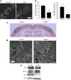
Synaptic activation reduces intraneuronal Aβ and protects against reductions in PSD-95 at synapses of APP mutant neurons. a, Western blot of cell lysates demonstrates reduced levels of intraneuronal Aβ, increased levels of βCTFs, and unchanged levels of full-length APP in g-LTP compared with control treated Tg2576 neurons. The ratio of Aβ normalized to APP was decreased 38 ± 11% in g-LTP-treated compared with control-treated Tg2576 neurons (n = 8; p < 0.01). The ratio of βCTFs normalized to APP was increased 93 ± 41% in g-LTP-treated compared with control-treated Tg2576 neurons (n = 13; p < 0.05). b, Aβ1-40 and Aβ1-42 ELISA of neuron lysates from g-LTP and controls (n = 6; p < 0.05). c, g-LTP also reduced levels of intraneuronal Aβ42 by 39 ± 4% compared with control-treated Tg2576 neurons, as determined by confocal immunofluorescence (n = 5; p < 0.01). d, immunofluorescence shows that g-LTP increased levels of PSD-95 puncta 35 ± 10% compared with control-treated Tg2576 neurons (n = 5; p < 0.01). Scale bars, 10 μm.
Figure 2.
Figure 2.
Synaptic activation reduces intraneuronal Aβ in hippocampal slices, whereas chronic inhibition of synaptic activity induces intraneuronal Aβ accumulation in vivo. a, Hippocampal slices prepared from Tg19959 mice and incubated for 8 h with KCl. Confocal microscopy of CA1 neurons demonstrates 13 ± 2% reduction of intraneuronal Aβ42 immunofluorescence in KCl-treated compared with control slices (n = 3; p < 0.01). b, Aβ1-42 ELISA of lysates from hippocampal slices treated with KCl versus control (n = 3; p < 0.05); Aβ1-40 ELISA of lysates from slices treated with KCl versus control (n = 3; p = 0.09). c, Representative section of Tg19959 brain stained for COX. The side corresponding to whisker removal (Surgery) shows a dramatic decrease of COX compared with the control side (Control). d, Confocal microscopy showing intraneuronal Aβ42 immunofluorescence of Tg19959 barrel cortex. The neuron cell bodies corresponding to the side with chronic synaptic inhibition (Surgery) show 10 ± 1% higher levels of Aβ42 compared with neurons on the control side (n = 4; p < 0.05). e, Metabolic labeling revealed decreased levels of newly generated Aβ (43 ± 11% decrease; n = 4; p < 0.01) and increased levels of newly generated βCTFs (22 ± 8% increase; n = 4; p < 0.05), in the presence compared with the absence of g-LTP. Aβ and βCTF values were normalized to newly generated APP. Levels of newly generated secreted Aβ (sAβ) (normalized to newly generated APP) were increased by 44 ± 15% in media of g-LTP activated compared with control Tg2576 neurons (n = 4; p < 0.05). Scale bars, 50 μm.
Figure 3.
Figure 3.
Alterations of synaptic proteins from extracellular Aβ1-42 occur via γ-secretase processing of APP. a, Treatment of wild-type neurons with Aβ1-42 increased levels of intraneuronal Aβ42 and reduced PSD-95 puncta as evident by immunofluorescence (central panels), but cotreatment with DAPT prevented these effects of Aβ1-42 (right panels). b, Quantitation of Aβ42 staining expressed as percentage of the Aβ42 immunofluorescence in the control (n = 5). c, Quantitation of PSD-95 expressed as a percentage of PSD-95 immunofluorescence of the control (n = 5). d, Treatment of wild-type neurons with Aβ1-42 reduced levels of surface NR1 as measured by surface biotinylation followed by Western blot. e, Quantitation of the ratio of surface NR1 to total NR1 in Aβ1-42 treated compared with untreated neurons and Aβ1-42 + DAPT treated compared with untreated neurons (n = 4). f, Treatment of APP KO neurons with Aβ1-42 or Aβ1-42 + DAPT showed unchanged levels of PSD-95 puncta as evident by immunofluorescence. **p < 0.01; scale bar: 10 μm.
Figure 4.
Figure 4.
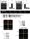
APP is transported to synapses with synaptic activity. a, Live cell imaging of Tg2576 cultured neurons transfected with APP-YFP. At steady state, movement of APP-YFP in dendrites is principally retrograde in these experimental conditions (see Materials and Methods). Kymographic analysis of APP-YFP moving vesicles (upper panel). Images were acquired at 10 s intervals for a total of 10 min. Arrowheads indicate movement of several APP-YFP containing vesicles. b, Quantitation of anterograde, retrograde and stationary APP-YFP containing-vesicles trafficking at steady state (n = 3). c, Live cell imaging of Tg2576 neurons transfected with APP-YFP after g-LTP. At steady state, movement of APP-YFP in neurites is predominantly anterograde. Images were acquired at intervals of 10 s for a total of 10 min. Arrowheads indicate movement of several APP-YFP containing vesicles. d, Quantitation of anterograde, retrograde and stationary APP-YFP containing-vesicles trafficking in Tg2576 dendrites with glycine treatment (n = 3). e, Representative images of APP-YFP vesicles before and after infusion with glycine. Images are shown 20 s apart. For complete movie with 10 s intervals see supplemental Movie S3, available at www.jneurosci.org as supplemental material. White arrows indicate two APP-YFP-containing vesicles that reverse direction from retrograde to anterograde on infusion of glycine at 20 s. Live cell images of Tg2576 cultured neurons transfected with APP-YFP were acquired at 10 s intervals for a total of 10 min per movie. The small black arrows indicate the time when glycine was added. Large black arrows indicate several other examples of reversal from retrograde to anterograde movement of APP-YFP containing vesicles in the kymograph. f, APP localizes to synapses on activation with g-LTP. Representative immunofluorescence for APP (green) and synapsin I (red) in Tg2576 neurons (upper panels). The lower panels demonstrate the increased localization (yellow) of APP with synapsin I on g-LTP. g, Levels of surface APP, normalized to total APP, were increased 89 ± 29% in g-LTP activated compared with control Tg2576 neurons, as measured by surface biotinylation followed by Western blot (n = 8, p < 0.05). h, Levels of surface APP, normalized to total APP, were reduced 33 ± 2% in synaptosomal preparations of g-LTP activated compared with untreated Tg2576 neurons, as assayed by surface biotinylation followed by Western blot (n = 5; p < 0.05). i, Surface APP (green) and synapsin I (red) immunofluorescence in g-LTP activated compared with control Tg2576 neurons in the absence of permeabilization. Compared with Figure 4 f (done with permeabilization), lower panels reveal no increase in localization of surface APP with synapsin I on synaptic activity. d, Distal; p, proximal; scale bar, 10 μm.
Figure 5.
Figure 5.
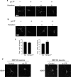
Involvement of neprilysin in g-LTP induced reduction of intraneuronal Aβ42. a, Fluorescent immunolabeling of Aβ40 in g-LTP activated versus nonactivated Tg2576 neurons in the presence or absence of the neprilysin inhibitor thiorphan. Despite inhibition of neprilysin, g-LTP reduces intraneuronal Aβ40 by 12 ± 1% as demonstrated by confocal immunofluorescence (n = 3; p < 0.01). b, Fluorescent immunolabeling for Aβ42 in g-LTP activated versus nonactivated Tg2576 neurons in the presence or absence of the neprilysin inhibitor thiorphan. Inhibition of neprilysin prevents g-LTP induced reduction of intraneuronal Aβ42, as demonstrated by confocal immunofluorescence (n = 3). c, Aβ1-40 and Aβ1-42 concentrations measured by ELISA in neuron lysates from g-LTP and controls in the presence of thiorphan. Inhibition of neprilysin allows for synaptic activity-induced reduction of Aβ1-40 but not of Aβ1-42 (n = 3; p < 0.05 for Aβ1-40 values). d, g-LTP activated NEP KO neurons and NEP KO control neurons. g-LTP induces reduction of intraneuronal Aβ40 (left panels; 39 ± 12% reduction; n = 6; p < 0.01) but not Aβ42 (right panels; n = 6) in NEP KO neurons, as determined by confocal immunofluorescence. Scale bars, 10 μm.

Similar articles

Cited by

References

    1. Almeida CG, Tampellini D, Takahashi RH, Greengard P, Lin MT, Snyder EM, Gouras GK. Beta-amyloid accumulation in APP mutant neurons reduces PSD-95 and GluR1 in synapses. Neurobiol Dis. 2005;20:187–198. - PubMed
    1. Almeida CG, Takahashi RH, Gouras GK. Beta-amyloid accumulation impairs multivesicular body sorting by inhibiting the ubiquitin-proteasome system. J Neurosci. 2006;26:4277–4288. - PMC - PubMed
    1. Ambrée O, Leimer U, Herring A, Görtz N, Sachser N, Heneka MT, Paulus W, Keyvani K. Reduction of amyloid angiopathy and Abeta plaque burden after enriched housing in TgCRND8 mice: involvement of multiple pathways. Am J Pathol. 2006;169:544–552. - PMC - PubMed
    1. Belinson H, Lev D, Masliah E, Michaelson DM. Activation of the amyloid cascade in apolipoprotein E4 transgenic mice induces lysosomal activation and neurodegeneration resulting in marked cognitive deficits. J Neurosci. 2008;28:4690–4701. - PMC - PubMed
    1. Billings LM, Oddo S, Green KN, McGaugh JL, LaFerla FM. Intraneuronal Abeta causes the onset of early Alzheimer's disease-related cognitive deficits in transgenic mice. Neuron. 2005;45:675–688. - PubMed

Publication types

MeSH terms

LinkOut - more resources