. 2009 Oct;83(20):10336-46.
doi: 10.1128/JVI.00747-09. Epub 2009 Aug 5.

Role for G-quadruplex RNA binding by Epstein-Barr virus nuclear antigen 1 in DNA replication and metaphase chromosome attachment

Affiliations

Role for G-quadruplex RNA binding by Epstein-Barr virus nuclear antigen 1 in DNA replication and metaphase chromosome attachment

Julie Norseen et al. J Virol. 2009 Oct.

Abstract

Latent infection by Epstein-Barr virus (EBV) requires both replication and maintenance of the viral genome. EBV nuclear antigen 1 (EBNA1) is a virus-encoded protein that is critical for the replication and maintenance of the genome during latency in proliferating cells. We have previously demonstrated that EBNA1 recruits the cellular origin recognition complex (ORC) through an RNA-dependent interaction with EBNA1 linking region 1 (LR1) and LR2. We now show that LR1 and LR2 bind to G-rich RNA that is predicted to form G-quadruplex structures. Several chemically distinct G-quadruplex-interacting drugs disrupted the interaction between EBNA1 and ORC. The G-quadruplex-interacting compound BRACO-19 inhibited EBNA1-dependent stimulation of viral DNA replication and preferentially blocked proliferation of EBV-positive cells relative to EBV-negative cell lines. BRACO-19 treatment also disrupted the ability of EBNA1 to tether to metaphase chromosomes, suggesting that maintenance function is also mediated through G-quadruplex recognition. These findings suggest that the EBNA1 replication and maintenance function uses a common G-quadruplex binding capacity of LR1 and LR2, which may be targetable by small-molecule inhibitors.


Figures
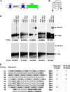
FIG. 1.
FIG. 1.
EBNA1 LR1 and LR2 bind to G-quadruplex RNA. (A) Schematic of EBNA1 protein. LR1 and LR2 are shown in blue, and the DBD is represented in green. (B) Representative image of a G-quadruplex structure. (C) The ability of EBNA1 LR1 and LR2 to bind to various RNA and DNA probes was determined by EMSA. Purified GST, GST-LR1, or GST-LR2 was incubated with 32P-labed probes and separated by electrophoresis on agarose gels. Bound and free probes are indicated by arrows. (D) List of probe sequences. All guanines that could potentially contribute to G-quadruplexes are in bold, and the ability of the probe to form a G-quadruplex is indicated. The results of LR1/LR2 EMSA are also indicated.
FIG. 2.
FIG. 2.
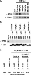
G-quadruplex-interacting compounds disrupt EBNA1 recruitment of ORC. (A) EBNA1 was immunoprecipitated from Raji cells, and the bead-bound complex was washed in the absence or presence of 10 μM G-quadruplex-interacting drugs (TMPyP3, TMPyP4, and BRACO-19) or a noninteracting analogue (TMPyP2) as indicated. Protein was eluted, separated by sodium dodecyl sulfate-polyacrylamide gel electrophoresis, and subjected to Western blotting with the indicated antibody. (B) Purified GST or GST-LR1 was bound to glutathione-Sepharose beads and incubated with HeLa nuclear extract. The bead-bound protein complex was washed in the absence or presence of 10 μM G-quadruplex-interacting drugs, as indicated. Associated proteins were determined as for panel A. (C) Purified GST or GST-LR1 was bound to glutathione-Sepharose beads and washed in the absence or presence of 10 μM BRACO-19, with or without 20 μg/ml RNase A, as indicated. The Sepharose beads were then blotted onto Whatman paper and dried. A digital image was taken, and the efficiency of BRACO-19 retention was scored as pigment intensity using ImageQuant. The percent BRACO-19 retention is indicated beneath each lane, normalized to the GST-LR1 control lane in the presence of BRACO-19.
FIG. 3.
FIG. 3.
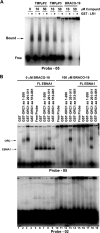
G-quadruplex drugs alter binding between EBNA1 and RNA (A) Binding between purified GST-LR1 and the 32P-labeled G-quadruplex probe RNA 05, in the absence or presence of 10 or 50 μM TMPyP2, TMPyP3, or BRACO-19, was measured by EMSA. The protein-bound probe is indicated by an arrow. (B) The ability of ORC1 peptides to bind to either G-quadruplex RNA or G-poor RNA was measured by EMSA. Purified GST, GST-ORC1 aa 1 to 200, GST-ORC1 aa 201 to 511, or GST-ORC1 aa 512 to 861 was incubated with either the 32P-labeled G-quadruplex probe RNA 05 or the 32P-labeled G-poor probe RNA 02 (lanes 1 to 4). Purified FL-EBNA1 was included with these reactions (lanes 5 to 9). The effect of 100 μM BRACO-19 on binding by these complexes was then tested (lanes 10 to 19). ORC1/RNA and EBNA1/RNA complexes are indicated by arrows.
FIG. 4.
FIG. 4.
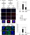
G-quadruplex drugs selectively kill EBV-positive cells and inhibit EBNA1-dependent replication at DS. (A) The copy number of the EBV genome was measured in EBV-positive Raji cells after 3 days of treatment, either mock (control) or 10 μM BRACO-19. Whole-cell DNA was extracted, and quantitative PCR with primers to either DS or β-actin was used to determine the relative ratio of EBV genome to cellular DNA. The unpaired t test was used to determine statistical significance between samples, and * denotes a two-tailed P value of less than 0.026. Error bars indicate standard deviations. (B) Reverse transcription-PCR (RT-PCR) analysis of mRNAs for EBNA1, EBNA2, EBNA3A, and LMP1 relative to cellular GAPDH mRNA in Raji cells treated with 0 or 10 μM BRACO-19 for 3 days. (C) The EBV-negative cell lines BJAB and DG75 and the EBV-positive cell lines Raji, LCL3456, and LCL3472 were cultured in the absence (control) or presence of 10 μM BRACO-19. After 6 days, cells were collected and stained with propidium iodide, and their cell cycle profile was analyzed by flow cytometry. Percentages of live cells are represented by the bar graph. The unpaired t test was used to determine statistical significance between samples and is indicated above the bars. Samples with a two-tailed P value of less than 0.007 are denoted by **, while samples with a two-tailed P value of less than 0.02 are denoted by *. (D) Schematic of constructs used in transient replication assay. The top protein schematic represents Flag-tagged FL-EBNA1. LR1 and LR2 are represented in blue, DBD in green, and the Flag tag in yellow. The middle protein schematic represents Flag-tagged DBD. The bottom protein schematic represents a Flag-tagged fusion peptide with 4×LR1-DBD. (E) Transient-replication assay. The constructs described in panel D were cotransfected into HeLa cells in the absence or presence of 10 μM BRACO-19, and DNA was harvested at 72 h posttransfection. DNA was either linearized with BamHI or digested with DpnI and separated by gel electrophoresis, and a reporter plasmid-specific probe was used to visualize replication. Percent replication was measured by PhosphorImager analysis and quantified by ImageQuant, with replication by FL-EBNA1 set at 100% replication (bottom left panel). (F) Samples of HeLa cells were also stained with propidium iodide (PI) and analyzed by flow cytometry to determine effects of BRACO-19 on cell cycle profile (bottom panel).
FIG. 5.
FIG. 5.
EBNA1 attachment to metaphase chromosomes is disrupted by G-quadruplex compounds (A) HeLa cells were transfected with either Cherry-EBNA1 (aa 1\ to 440) (denoted as Cherry-EBNA1Δ) or Cherry vector alone, sorted for red fluorescent protein (RFP) signal, replated, and then treated with 0 or 10 μM BRACO-19. The experimental design is shown in the schematic. Representative images of DNA staining (blue), Cherry protein localization (red), or the merge image are shown. Beneath each column a merge image of one of the metaphase spreads from the field is enlarged for detail. (B) Percentage of nuclei that scored as being positive for Cherry protein colocalization to metaphase spreads. n represents the number of metaphase spreads scored for each condition. The unpaired t test was used to determine statistical significance between treated and untreated Cherry-EBNA1 and is indicated above the bars. Samples with a two-tailed P value of 0.0001 are denoted by **. Error bars indicate standard deviations. (C) To ensure that 10 μM BRACO-19 did not alter Cherry-EBNA1 expression, protein samples were collected and examined with antibody against either EBNA1 or β-actin by Western blotting. (D) GFP-FL-EBNA1 attachment to metaphase chromosomes in the presence of 0 or 10 μM BRACO-19 was also examined. HeLa cells were transfected with either GFP vector or GFP-FL-EBNA1. Metaphase spreads were examined by epifluorescence for GFP association with metaphase chromosomes. Representative metaphase spreads showing the merged image of DNA (blue) and GFP protein colocalization (green) have been enlarged for detail. (E) Quantification of nuclei from the experiment shown in panel D. n represents the number of metaphase spreads scored for each condition. The unpaired t test was used to determine statistical significance between treated and untreated GFP-FL-EBNA1 and is indicated above the bars. Samples with a two-tailed P value of 0.0001 are denoted by **.

Similar articles

Cited by

References

    1. Altmann, M., D. Pich, R. Ruiss, J. Wang, B. Sugden, and W. Hammerschmidt. 2006. Transcriptional activation by EBV nuclear antigen 1 is essential for the expression of EBV's transforming genes. Proc. Natl. Acad. Sci. USA 103:14188-14193. - PMC - PubMed
    1. Atanasiu, C., Z. Deng, A. Wiedmer, J. Norseen, and P. M. Lieberman. 2006. ORC binding to TRF2 stimulates OriP replication. EMBO Rep. 7:716-721. - PMC - PubMed
    1. Avolio-Hunter, T. M., and L. Frappier. 1998. Mechanistic studies on the DNA linking activity of Epstein-Barr nuclear antigen 1. Nucleic Acids Res. 26:4462-4470. - PMC - PubMed
    1. Bashaw, J. M., and J. L. Yates. 2001. Replication from oriP of Epstein-Barr virus requires exact spacing of two bound dimers of EBNA1 which bend DNA. J. Virol. 75:10603-10611. - PMC - PubMed
    1. Bochkarev, A., J. A. Barwell, R. A. Pfuetzner, E. Bochkareva, L. Frappier, and A. M. Edwards. 1996. Crystal structure of the DNA-binding domain of the Epstein-Barr virus origin-binding protein, EBNA1, bound to DNA. Cell 84:791-800. - PubMed

Publication types

LinkOut - more resources